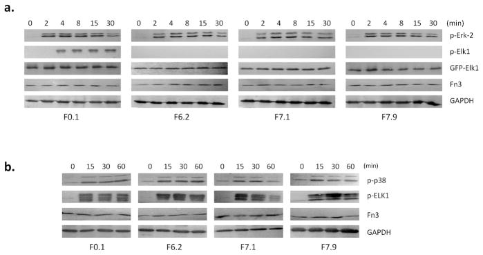
Figure 4. Activity of engineered Fn3 in HEK293 cells.

a. HEK293 cells were transiently transfected with pCMVFn3-IEGFP-Elk1(307-428) and stimulated with EGF to activate the ERK-2 pathway. in the resulting Elk1 phosphorylation was inhibited by F6.2, F7.1, and F7.9. The stable expression of EGFP-Elk1 was checked by fluorescence microscopy and by Western blot with anti-GFP antibody (Fig. S7). Fn3 expression was checked using 9E10 against the cMyc tag. The number of cells used in the analysis was normalized using GAPDH.
b. HEK293 cells expressing various Fn3 clones were stimulated with 100 mM NaCl to activate the p38 pathway. The monobodies had little or no effect on the p38-dependent phosphorylation of Elk1.
