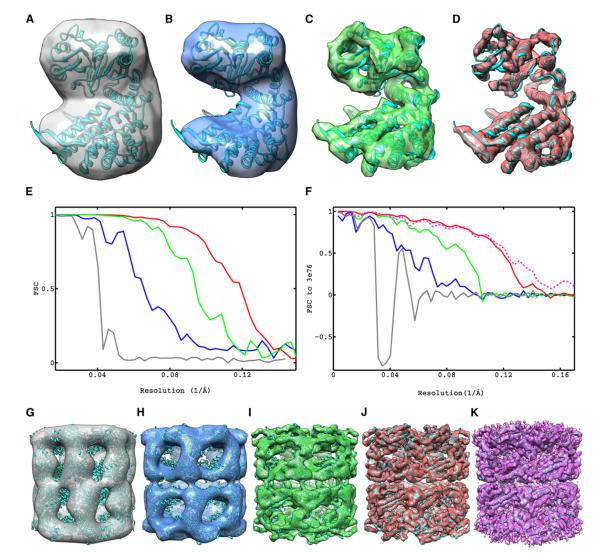
Figure 4. Progressive Improvement in Resolution Achieved by Each Component of Constrained Single-Particle Tomography and Comparison to Highest Resolution Map of GroEL Reported Using Cryo-EM.
Maps are represented as iso-surfaces with the fitted X-ray coordinates.
(A) Reconstruction by conventional subvolume averaging.
(B–D) Fourier-based CTF-corrected reconstructions using only first 11 exposures in the tilt series and alignments from subvolume averaging (B), after traditional projection-matching refinement of image shifts (C), and after constrained projection-matching refinement (D).
(E) FSC plots of maps in (A)–(D) obtained from the correlation of reconstructions between random halves of the image data set, indicating resolutions measured by the 0.5 cutoff criteria of 24.5, 15.3, 10.6, and 8.4 Å, respectively.
(F) FSC plots against a map derived from the X-ray model indicating resolutions measured by the 0.5 cutoff criteria of 34.1, 18.2, 10.8, and 8.5 Å for maps in (A)–(D) and 8.2 Å for map shown in (K).
(G–J) Maps of the entire complex corresponding to the subunits shown in (A)–(D).
(K) Map (4.2 Å) of GroEL (EMDB ID 5001) obtained by traditional single-particle cryo-EM (Ludtke et al., 2008), using 20,401 particles, 25–36 e−/Å2 and 300 kV imaging (iso-surface shown at suggested contour level of 0.597).

