Figure 1.

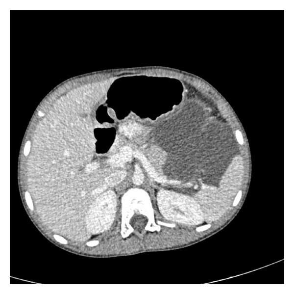
Contrast-enhanced CT axial scan shows retroperitoneal lobulated-septated cystic mass between spleen, stomach, and pancreas. Splenic vein and artery borders are in the cystic mass. Also cystic mass reaches to the pararenal space.

Contrast-enhanced CT axial scan shows retroperitoneal lobulated-septated cystic mass between spleen, stomach, and pancreas. Splenic vein and artery borders are in the cystic mass. Also cystic mass reaches to the pararenal space.