
Figure 1. Comparison of early myocardial development in chick, mouse, and zebrafish.
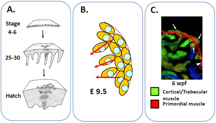
A. Clonal analysis of cardiomyocyte precursor in chick by single cell retroviral tagging. B. Expansion of murine myocardial precursors cells at embryonic day 9.5. C. Development of cortical myocardial layer from trabecular cardiomyocytes in the zebrafish heart. Arrows represent cortical cardiomyocytes and their putative trabecular origin. Adapted with permission from references #1, 3, and 5.