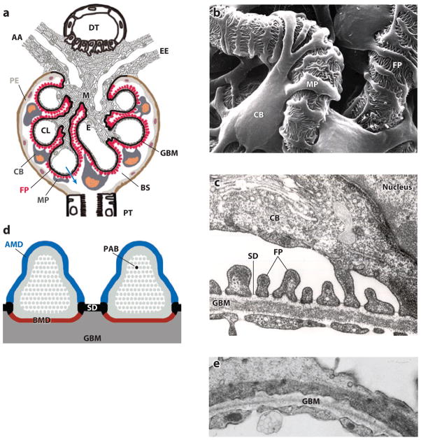
Figure 1.
The function of podocytes is based on their intricate cell architecture. (a) A glomerulus contains a capillary tuft that receives primary structural support from the glomerular basement membrane (GBM). Glomerular endothelial cells (E) lining the capillary lumen (CL) and mesangial cells (M) are located on the blood side of the GBM, whereas podocyte foot processes (FP) cover the outer part of the GBM. Podocyte cell bodies (CB) and major processes (MP) float in Bowman’s space (BS) in primary urine. Along its route from the CL to BS (blue arrow), the plasma ultrafiltrate passes through the fenestrated endothelium, the GBM, and the filtration slits that are covered by the slit diaphragm (SD). AA, afferent arteriole; DT, distal tubule; EE, efferent arteriole; PE, parietal epithelium; PT, proximal tubule. (b) Scanning electron microscopy view from BS highlighting the intricate shape of podocytes. MP link the CB to FP, which interdigitate with FP of neighboring podocytes, thereby forming the filtration slits. (c) Transmission electron microscopy image of the filtration barrier consisting of fenestrated endothelium, GBM, and podocyte FP with the SD covering the filtration slits. (d) Podocyte FP are defined by three membrane domains: the apical membrane domain (AMD) (blue), the basal membrane domain (BMD) (red), and the SD (black). All three domains are connected to the underpinning actin cytoskeleton (gray) and with each other. PAB denotes a single parallel, contractile actin bundle containing α-actinin-4, myosin 9, and synaptopodin. (e) Under conditions of proteinuria, FP lose their normal interdigitating pattern and instead show effacement. A continuous sheet of cytoplasm filled with reorganized actin filaments is clearly visible.

