Fig. 2.
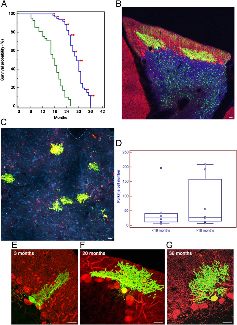
Developing mouse cerebellar neurons transplanted in utero into the developing rat CNS integrate and survive to the rat host maximum lifespan, doubling the average lifespan of the neurons in the donor mice strain. (A) Kaplan–Meier plot showing lifespan of siblings of donor mice and rat host. Rats perfused electively were excluded from the survival curve. Each red dot indicates a transplanted rat in which we found mouse cells. (B) Parasagittal section from confocal microscopy of a 20-mo-old rat cerebellum. Fluorescences are as follows: EGFP (green), anti-calbindin antibody (red), overlapping fluorescence (yellow), and DAPI nuclear staining (blue). Two grafted mouse PCs are integrated in the host PC and molecular layers, and granular cells of mouse origin are visible in the granular layer (asterisks). (Scale bar: 25 μm.) (C) Same as Fig. 1A, but coronal section of the brainstem and fourth ventricle of a 36-mo-old graft, in which six ectopic calbindin/EGFP-positive PCs are visible in the brainstem outside the dorsal cochlear nucleus. Interrupted line indicates the ventricle. (Scale bar: 50 μm.) (D) Plot of the number of PCs found in the transplanted animals at different ages (<19 mo, rats perfused before 19 mo of age; >18 mo, rats perfused after 18 mo to 36 mo of age). Each square represents a single animal: <19 mo, n = 6; >18 mo, n = 7. Differences in mean and median number of PCs in the two groups were not significant (P = 0.5968, unpaired Student t test; P = 0.5338, Mann–Whitney test). Horizontal line indicates the median; central box encloses values from the lower to upper quartile; vertical line extends from the minimum to the maximum value, excluding outside and far out values. (E–G) Parasagittal sections as in Fig. 1A but without DAPI. Rat cerebella at 3, 20, and 36 mo of age showing mouse-derived PCs. The span of mouse PC dendrite did not reach the top of the host molecular layer and remained approximately the same at all survival times. (Scale bar: 25 μm.)

