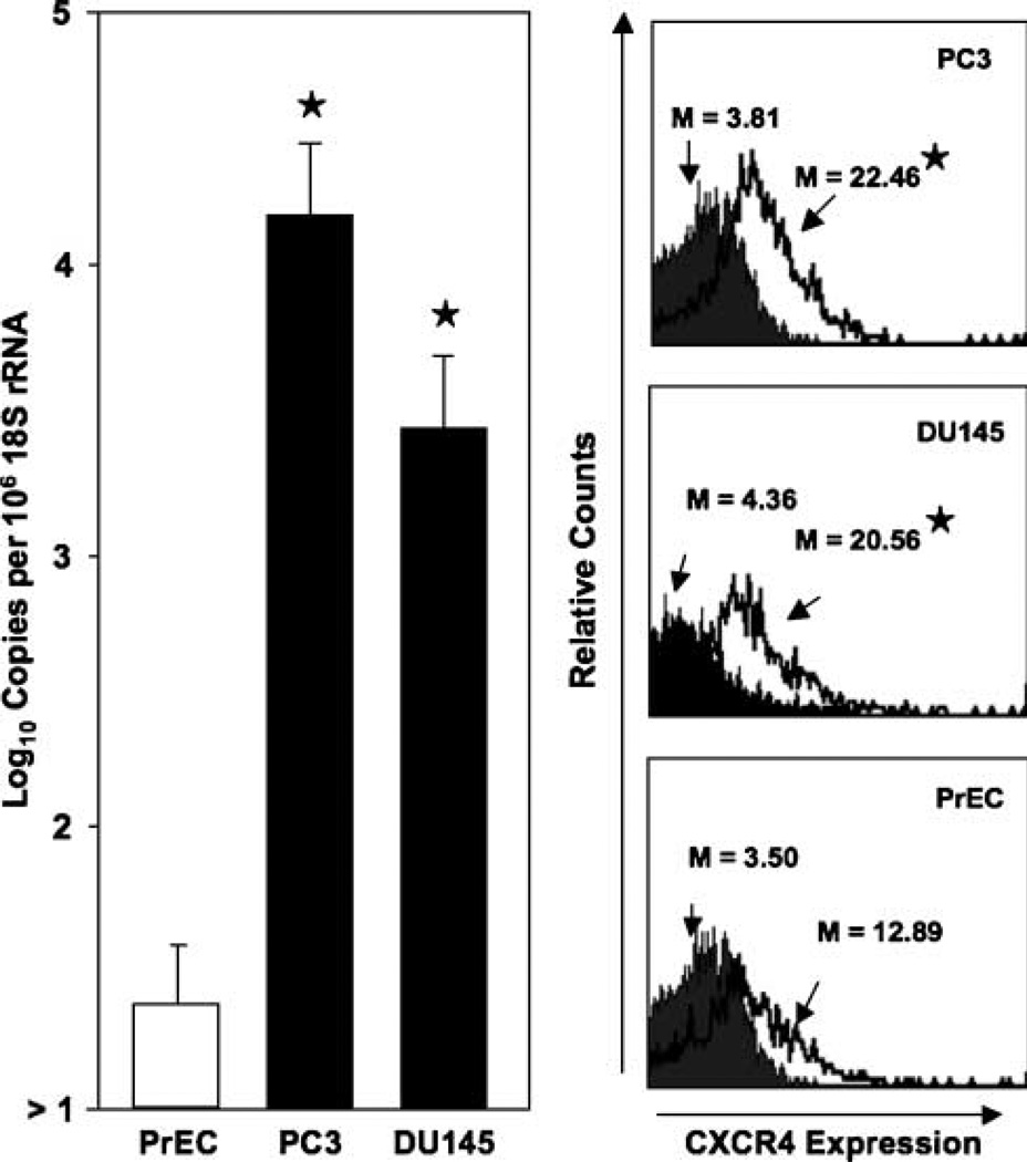
Figure 1.
CXCR4 expressed by prostate cancer cell lines and PrEC cells. Total RNA was isolated from prostate cancer cell lines and PrEC cells and quantitative real-time PCR analysis of CXCR4 mRNA expression was done in triplicate. Copies of transcripts were expressed relative to actual copies of 18S rRNA ± SE. PC3 and DU145 cell lines and PrEC cells were stained with PE-conjugated anti-CXCR4 (open histogram) or PE-conjugated isotype control antibodies (solid histogram) and quantified in triplicate by flow cytometry. M, mean fluorescent intensities of CXCR4-positive cells. ★, P < 0.001, statistical significance between normal and prostate cancer cell lines.

