
Figure 5. Immunohistology.
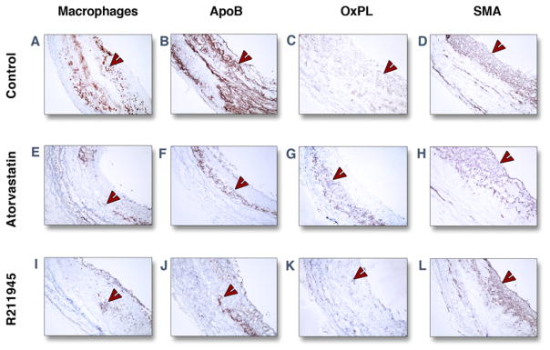
Representative immunohistological images of individual groups of the abdominal aorta at 3 months. Staining (red-brown, red arrowheads) for macrophages (A, E, I), apolipoprotein B (ApoB) (B, F, J), oxidized phospholipids (OxPL) (C, G, K), and smooth muscle actin (SMA) (D, H, L) are shown. Objective magnification: ×10. The lumen is on the right-hand side from the tissue.