
Figure 1.
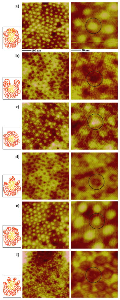
AFM panels of the unique PS-b-P4VP nanostructures monitored during time-dependent exposure to CHCl3 vapor; a) original spheres, b) holes, c) reformed spheres, d) embedded spheres, e) enlarged spheres and f) cylinder precursors. AFM images clearly demonstrate the periodic two-dimensional arrays consisting of the well-defined nanostructures at each time period of annealing for a) 0 h, b) 3 h, c) 9 h, d) 16 h, e) 25 h, and f) 29 h. The scan sizes of the images shown on the left and right column are 500 × 500 nm and 140 × 140 nm, respectively. A dark circle is inserted in the zoomed-in AFM panels in order to highlight each, independently addressable, PS-b-P4VP unit in its hexagonally packed configuration. As a guide, illustrations showing possible polymeric chain distributions in each micelle are inserted next to each AFM panel for each time-dependent PS-b-P4VP nanostructure. Red and yellow chains in the cartoons represent PS and P4VP, respectively.