
Fig. 5.
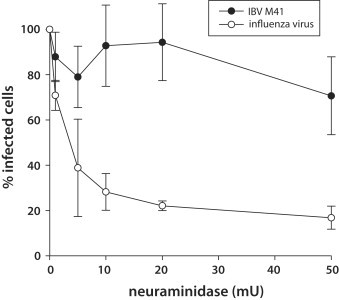
Effect of neuraminidase treatment on 3T3–DC-SIGN cells infected by IBV-M41 3T3–DC-SIGN cells were pre-treated with neuraminidase and then were infected by M41 (103 TCID50/ml), or a control virus, influenza A/WSN/33 (MOI of 5). At 12 h post infection (M41) or 5 h post infection (WSN), cells were fixed and stained for immunofluorescence microscopy with 15:88 anti-S1 mouse monoclonal antibody (M41) or anti-NP mouse monoclonal antibody (WSN/33). Cells were quantified by scoring the percentage of cells positive for viral antigen. >200 cells were quantified from three independent experiments. Error bars represent the standard deviation from the mean.