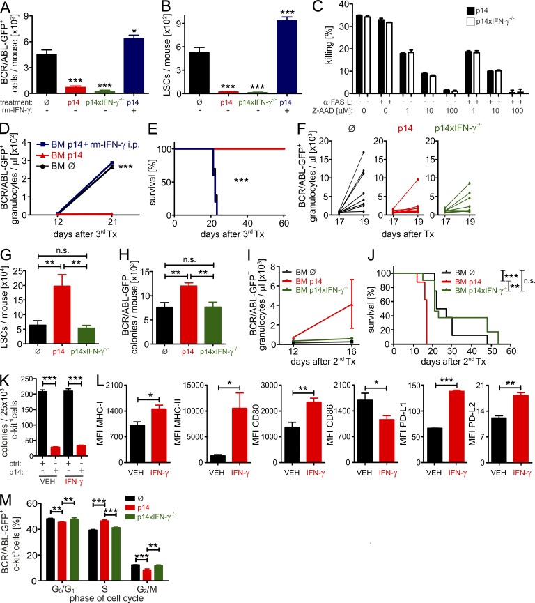
Figure 6.
CTL-secreted IFN-γ increases LSC numbers. (A) 2 × 10⁴ H8 LSCs were FACS-purified 20 d after transplantation and were transferred i.v. into irradiated (6.5 Gy) secondary BL/6 recipients. 18 h later, mice were either left untreated (Ø, n = 11) or treated with 3 × 10⁶ FACS-purified p14 (n = 12) or p14xIFNγ−/− (n = 3) CTLs i.v. One group of mice received p14 CTLs and was treated with 2.5 × 10⁵ U of rm-IFN-γ twice daily for two consecutive days (n = 5). After 48 h, BM was harvested and analyzed for BCR/ABL-GFP+ (A) cells and LSCs (B) by FACS. Pooled data from two independent experiments are shown. (C) Duplicates of bulk BM cells from H8 CML mice were either incubated alone or were co-cultured with CTLs at a ratio of 5:1 with or without 10 µg/ml anti–FAS-ligand, in the presence or absence of the indicated concentrations of the granzyme B inhibitor Z-AAD-CMK. After 4 h, the killing of BCR/ABL-GFP+ cells was determined. (D and E) 3 × 10⁶ BM cells from p14-treated (red line, n = 5), p14+rm-IFN-γ–treated (blue line, n = 5), or untreated (black line, n = 10) H8 CML mice were tertiarily transplanted into irradiated (6.5 Gy) BL/6 mice. Numbers of BCR/ABL-GFP+ granulocytes/µl blood (D) and Kaplan-Meier survival curves (E) resulting from tertiary transplantations are shown. (F) BCR/ABL-GFP+ granulocytes in blood of primary H8 CML mice either left untreated (Ø) or treated with 3 × 10⁶ FACS-purified p14 CTLs or p14xIFN-γ−/− CTLs (n = 8 each). LSC numbers (G) and BCR/ABL-GFP+ colonies per mouse (H) 2 d after adoptive transfer. (I–J) 3 × 10⁶ BM cells from primary H8 CML mice left untreated (black line, n = 8) or treated with p14 CTLs (red line, n = 8) or p14xIFN-γ−/− CTLs (green line, n = 11) were secondarily transplanted into irradiated (6.5 Gy) BL/6 mice. (I) Numbers of BCR/ABL+ granulocytes/µl blood and (J) Kaplan-Meier survival curves resulting from secondary transplantations. (K) H8 CML mice were treated with either vehicle (VEH, n = 5) or 2.5 × 10⁵ U of rm-IFN-γ (n = 5) i.p. twice daily on two consecutive days (d17 and d18 after primary transplantation). 2.5 × 10⁴ FACS-purified H8 BCR/ABL-GFP+ c-kithi cells from VEH- or IFN-γ–treated H8 CML mice were co-incubated with 2.5 × 10⁴ naive BL/6 CD8+ T cells (ctrl) or p14 CTLs (p14) overnight, followed by transfer into methylcellulose in duplicates. Colonies were enumerated 7 d later. (L) Mean fluorescent intensity (MFI) of MHC class I, MHC class II, CD80, CD86, PD-L1 and PD-L2 on LSCs from VEH- or IFN-γ–treated H8 CML mice. (M) Cell cycle analysis by DAPI stainings of total c-kithi cells from primary H8 CML mice left untreated (Ø, n = 6) or treated with p14 (n = 5) or p14xIFN-γ−/− (n = 5) CTLs. Data are displayed as mean ± SEM. Statistics: one-way ANOVA (A, B, G–H, and M), two-way ANOVA (D and I), Student’s t test (K and L), and log-rank test (E and J). *, P < 0.05; **, P < 0.005; ***, P < 0.0005.

