
Fig. 3.
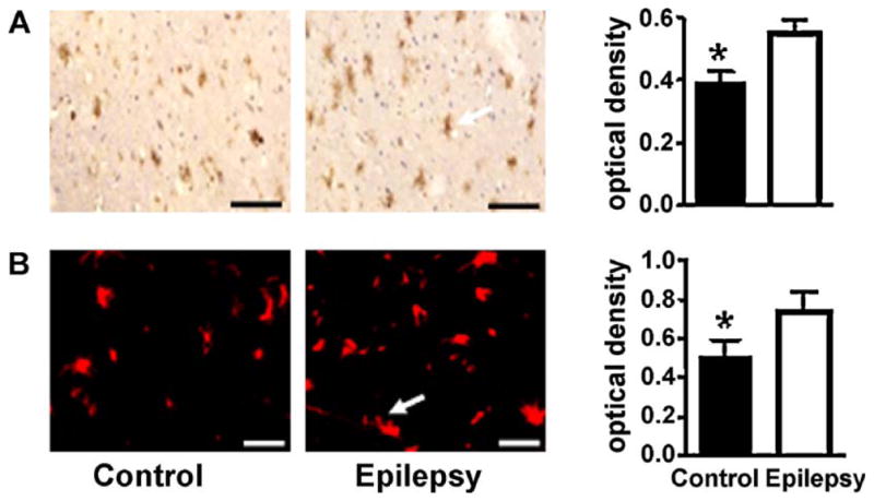
GFAP immunoreactivity in the temporal lobe of IE patients by immunohistochemical staining. (A) Faint positive staining in the control illustrates decreased GFAP expression. Strongly positive staining in the cortex of the temporal lobe of a patient with IE reflects increased GFAP expression (buffy particles in the cytoplasm of glial cells). (B) Immunofluorescence of μ-calpain expression in the control and patients with temporal lobe epilepsy. The white arrows indicate positive cells (glial cell) representing increased μ-calpain expression. Scale bars = 75 μm. The data are expressed as mean ± SD, and the value of *p < 0.05 is indicated as significantly different.