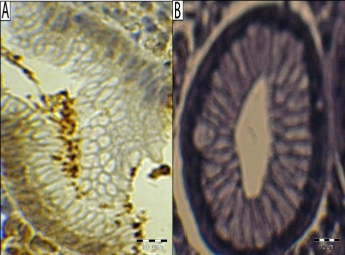
Fig. 1.
Immunohistological demonstration of H. pylori. (A) Clusters of immunolabeled organisms at the surface of pyloric mucosa of a patient infected with Helicobacter pylori, immunohistochemical staining for H. pylori (primary antibody: Bo471 DAKO, Denmark), hematoxylin counterstain, positive control reaction; (B) negative control reaction. Bar line = 10 µm

