
Figure 2.
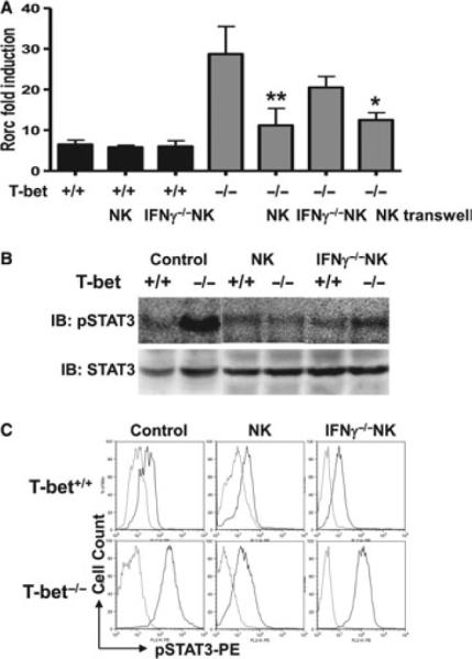
Loss of T-bet renders Rorc and STAT3 hyperresponsive to activation signals provided by NK cells. CD4+ T cells were sorted from pooled spleen and lymph node cells of acetylcholine receptor (AChR)-primed WT and T-bet−/− mice. A portion of these wells also received NK cells. Parallel co-cultures were set up using transwells. (A) Fold induction of Rorc transcripts at 3 days after stimulation measured by qRT-PCR. (B) STAT3 phosphorylation (pSTAT3) analysed by Western blotting. (C) pSTAT3 in AChR-stimulated CD4+ T cells measured by FACS by intracellular staining. Data represent two independent experiments.