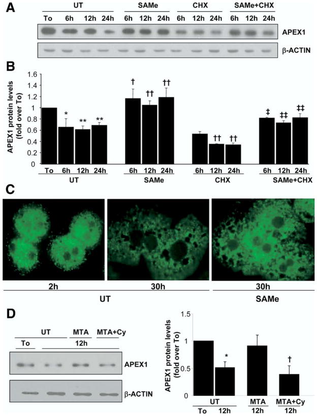
Figure 5.
Effect of SAMe and MTA treatment on APEX1 protein levels in mouse cultured hepatocytes. Mouse hepatocytes were treated with SAMe (2 mmol/L) or MTA (1 mmol/L) for different time points. When indicated, cells were pretreated with cycloheximide (CHX, 5 μg/mL) for 5 minutes or cycloleucine (Cy, 20 mmol/L) for 1 hour. (A) At the indicated times, Western blot analysis was done for APEX1 or β-actin. (B) Results are expressed as fold over To cells (mean ± SEM) from 3 to 5 independent experiments. *P < .005 vs To, **P < .001 vs To, †P < .05 vs UT at respective time points, ††P < .02 vs UT at respective time points, ‡P < .05 vs CHX at respective time points, ‡‡P < .02 vs CHX at respective time points. (C) Subcellular localization of APEX1 was evaluated by confocal microscopy as described in the Materials and Methods section. APEX1 protein appears green on fluorescent microscopy. (D) Representative Western blots are shown from 3 independent experiments showing the effect of MTA with or without Cy pretreatment. Densitometric changes are shown and expressed as fold over To in the graph on right. *P < .002 vs UT, †P < .002 vs MTA.

