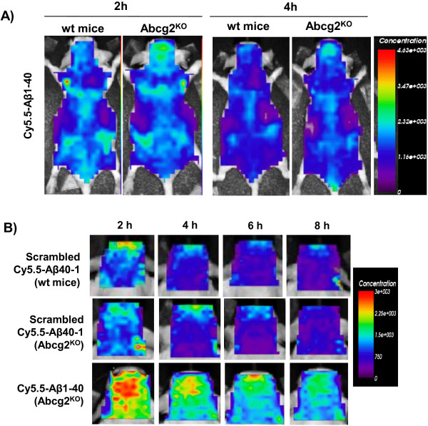
Figure 3.
Serial concentration images of Abcg2-KO and wild-type mice injected i.v. with either Cy5.5-labeled scrambled Aβ40-1 or Aβ1-40peptides. The peptides (100 μg in 200 μL volume) were injected i.v. and whole body and head ROIs of animals were imaged at 2, 4, 6, and 8 h using the eXplore Optix 670. Panel A shows the whole body (dorsal) images of wild-type and Abcg2-KO mice 2 and 4 h after i.v. injection of Cy5.5- Aβ1-40. Panel B shows head ROI fluorescence concentration images over time in wild-type mice injected with scrambled Aβ40-1, and Abcg2-KO mice injected with either Cy5.5-labeled scrambled Aβ40-1 or with Cy5.5-labeled Aβ1-40 peptide. The images shown were analyzed with ART Optix Optiview software and are representative of 4 animals per group.

