
Figure 5.
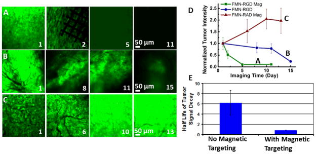
RGD-conjugated FMN in combination with external magnetic control expedites tumor regression in a U87MG human glioblastoma xenograft mouse model. (A–C) EGFP-transfected tumor image channels show tumor intensity change within days of imaging for FMN-RGD together with magnetic targeting (A), FMN-RGD without magnetic targeting (B), and FMN-RAD under magnetic targeting (C). Day 1 is the day of FMN injection and magnetic targeting. The permanent magnet was placed for 2 hours, and the Ni micromesh was removed the following day to better observe the imaging area. (D) The normalized tumor intensity vs. imaging time curves demonstrate that FMN-RGD together with magnetic targeting can expedite tumor regression (n=3, P<0.05). The fluorescence image intensity scales were set so that the brightest image within each series (A–C) was near saturation. (E) The half-lives of tumor signal decay demonstrate significantly faster tumor regression for FMN-RGD injection with magnetic targeting (n=3, 0.853 days) compared to without magnetic targeting (n=3, 6.197 days). The half-lives were obtained by fitting each of the six FMN-RGD injection curves (Figure 4D and Supplementary Figure 3) to a first order exponential decay function.