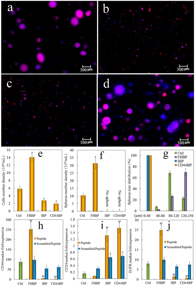
Figure 6. Comparison of tumorsphere formation in PEGDA gels conjugated with CD44BP, IBP, or FHBP.
Representative fluorescent images of the tumorsphere size and distribution for 4T1 cells encapsulated in PEGDA gels (1.4×10⁵ cells/ml) without peptide conjugation (a), conjugation with CD44BP (CD44BP, b), conjugation with RGD integrin-binding peptide (IBP, c) and conjugation with fibronectin-derived binding peptide (FHBP, d) and cultured in the stem cell culture medium for 9 days. Effect of cell binding peptide on cell number density (e), tumorsphere number density (f) and sphere size distribution (g) for 4T1 tumor cells encapsulated in PEGDA gel and incubated in the stem cell culture medium for 9 days. Effect of cell binding peptide on CD44 (h), CD24 (i) and EGFR (j) mRNA marker expression for 4T1 tumor cells encapsulated in PEGDA hydrogel and incubated in the stem cell culture medium for 9 days. RNA levels of the cells were normalized to those at time zero. A star indicates a statistically significant difference (p<0.05) between the test group and “Ctrl”. Two stars indicates a significant difference (p<0.05) between the wild type and scrambled peptides for the same conjugated peptide. Values are expressed as mean ± SD (n = 3).

