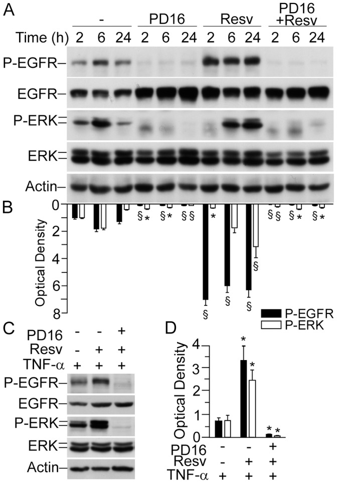
Figure 4. Abrogation of both constitutive and resveratrol-dependent EGFR and ERK phosphorylation by specific inhibitor of EGFR kinase.

(A) Western blot analysis of whole-cell lysates. Keratinocytes were incubated with 2 µM PD168393 (PD16) for 30 minutes prior to addition of 50 µM resveratrol (Resv) for the indicated time-points. (B) Quantification of Western blot bands by densitometry. *P<0.05 and § P<0.01 versus untreated controls for each time-point. (C) Western blot analysis of whole-cell lysates. Keratinocytes were incubated with 2 µM PD16 prior to addition of 50 µM Rv for 1 h. Subsequently, cells were further treated for 12 h with 50 ng/ml TNFα. (D) Quantification of Western blot bands by densitometry. *P<0.05 versus controls treated with TNFα only. Data are representative of three independent experiments.
