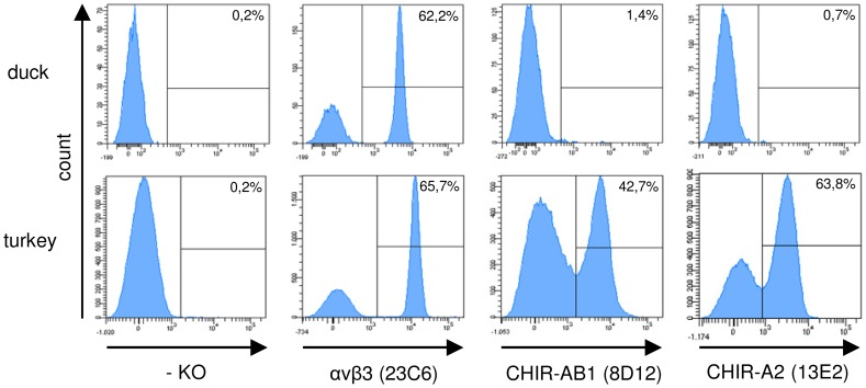
Figure 5. Immunofluorescence staining of duck and turkey PBMC.
Blood was separated via density centrifugation and stained with mab against αVβ3 and various CHIR followed by secondary antibody conjugates. Histograms of one typical out of four separate experiments is shown and frequencies of positive cells are indicated.

