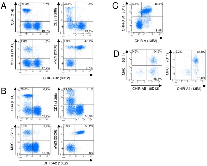
Figure 6. Double fluorescence analyses of turkey PBMC.
Cells were labeled with either CHIR-AB1 specific mab 8D12 (A) or CHIR-A2 specific mab 13E2 (B) in combination with the T-cell specific mab CD4 and CD8, the class II specific mab 2G11 and the αVβ3 specific mab 23C6. (C) Double staining of blood leukocytes with 8D12 and 13E2. Gates were set according to forward and sideward scatter features on the lymphocyte gate in A, B and C. (D) Combined immunfluorescence of 8D12 or 13E2 in combination with the MHC class II specific mab 2G11 as in (A) or (B), but gated on the larger monocytes based on forward/side scatter characteristics. Frequencies of positive cells are indicated in the quadrants. One representative of three experiments is shown.

