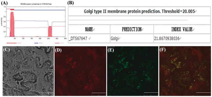
Figure 3. Subcellular localization of GhGalT1 protein.
(A) The prediction of trans-membrane helices in GhGalT1 protein. GhGalT1 belongs to type II membrane proteins with an obvious single trans-membrane helice as predicted by the TMHMM2.0 program. (B) GhGalT1 protein with index values greater than the threshold is predicted as Golgi protein. (C) to (F) Fluorescent protein-tagged GhGalT1 fusion proteins were coexpressed with GONST1-YFP (Golgi apparatus marker) into the abaxial side of a young tobacco (Nicotiana tabacum) leaf epidermis. The signals were visualized with a laser confocal microscope. (C) Bright field photograph of leaf epidermal cells of tobacco (Nicotiana tabacum). (D) The image of GONST1-YFP (Golgi apparatus marker) fluorescence. (E) The image of GhGalT1-GFP fluorescence. (F) Image (E) was merged with image (D). Scale bar = 50 µm.

