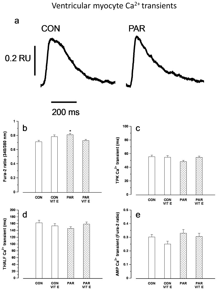
Figure 3.
Typical records of Ca2+ transients in myocytes from control and PAR treated rats (a). Resting fura-2 ratio (b), time to peak (TPK) Ca2+ transient (c), time to half (THALF) decay of the Ca2+ transient (d) and amplitude of the Ca2+ transient (e). Data are mean±SEM, n = 20–32 cells from 3 hearts in each treatment group. CON = Control, PAR = Paraquat, VIT E = Vitamin E.

