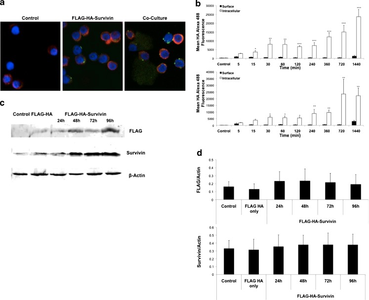
Fig. 1.
Normal T cells take up tumor-released Survivin. a Normal T cells were isolated by magnetic separation and cultured with FLAG-HA-Survivin or co-cultured with POZn-WT-Survivin cells for 24 h. Confocal images show uptake and surface binding of Survivin using a 60x oil immersion objective lens (Red – Actin, Green – FLAG-HA-Survivin, blue – DAPI). Representative of 2 independent experiments. b Surface and intracellular mean fluorescence intensity of HA in CD4+ (top) and CD8+ (bottom) T cells over time. Representative of 3 independent experiments. (Mean ± SD for 3 independent experiments; *p ≤ 0.05, **p ≤ 0.01, ***p ≤ 0.001 Intracellular vs. Surface). c Western blotting of T cells shows the presence of intracellular Survivin and FLAG tagged Survivin over time. Blots are representative of 3 independent experiments. d Densitometry shows Mean ± SD for 3 independent experiments, 2 blots each, n = 6

