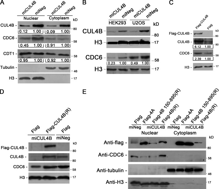
Figure 2.
The levels of CUL4B and nuclear CDC6 are positively correlated. (A) miCUL4B and miNeg HeLa cells were separated into nuclear and cytoplasmic fractions. The protein levels were determined with indicated antibodies. (B) Nuclear proteins prepared from stable CUL4B RNAi or negative control HEK293 and U2OS cells were immunoblotted with indicated antibodies. (C) Nuclear proteins prepared from 293T cells transiently transfected with indicated constructs were analyzed by Western blot. Band intensity given underneath gel images in A–C was measured using ImageJ software (NIH, Bethesda, MD), presented as fold change. (D) miCUL4B and miNeg HeLa cells were transfected with indicated plasmids. At 72 h after transfection, nuclear proteins were extracted and analyzed by Western blot. (E) miCUL4B and miNeg HeLa cells transfected with equal amounts of the indicated plasmids were separated into nuclear and cytoplasmic fractions and the protein expression was analyzed by Western blot.

