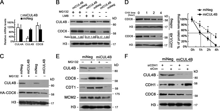
Figure 4.
Proteolysis of CDC6 is accelerated in CUL4B knockdown cells. (A) The mRNA levels of CUL4A, CUL4B, and CDC6 in miCUL4B and miNeg HeLa cells were measured by real-time PCR. The normalized expression in miNeg cells was set as 1. The assay was performed in triplicate, and relative means ± SE are shown. (B) miCUL4B and miNeg HeLa cells were treated or untreated with 30 µM MG132 for 3 h or 10 µM LMB for 5 h and nuclear proteins were extracted and analyzed by Western blot. Band intensity given underneath gel image was measured using ImageJ software, presented as fold change compared with control cells. (C) miCUL4B and miNeg HeLa cells were transiently transfected with HA-tagged CDC6 construct. 72 h after transfection, cells were treated or untreated with 30 µM MG132 for 3 h and nuclear proteins were analyzed by Western blot. (D) miCUL4B and miNeg HeLa cells were treated with 50 µg/ml cycloheximide and harvested at the indicated time points. Equal amounts of nuclear proteins were analyzed by Western blot. The nuclear CDC6 proteins expression was quantified by densitometric analysis using ImageJ. Expression is represented as the percentage remaining relative to time zero; data are expressed as means ± SEM (n = 3). *, P < 0.05 vs. miNeg. (E) Western blot analyses of indicated protein levels in the chromatin-bound fraction prepared from miCUL4B and miNeg HeLa cells treated or untreated with 30 µM MG132 for 3 h. (F) miCUL4B and miNeg HeLa cells were transfected with indicated siRNAs. 48 h after transfection, cells were treated with 0.5 mM mimosine for 22 h and nuclear proteins were analyzed by Western blot.

