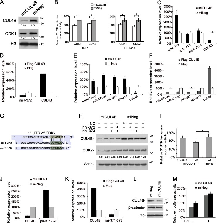
Figure 6.
CUL4B up-regulates CDK2 expression by repressing miRNA. (A) Nuclear proteins prepared from miCUL4B and miNeg HeLa cells were analyzed by Western blot. (B) Luciferase reporter assay of miCUL4B and miNeg cells transiently transfected with pmir-GLO-CDK1-3′UTR, pmir-GLO-CDK2-3′UTR, and pmir-GLO empty vector. Values were corrected for the expression of Renilla luciferase and compared with that of pmir-GLO. Levels of luciferase activity were compared with those of miNeg-transfected cells, normalized to 1. The results are given as mean values from three independent experiments. Bars represent means ± SD. *, P < 0.05 vs. miNeg. (C) miR-372 expression is negatively regulated by CUL4B. The levels of indicated miRNAs in miCUL4B and miNeg HeLa cells were measured by real-time PCR. The normalized expression in miNeg cells was set as 1. The assay was performed in triplicate, and relative means ± SE are shown. (D) The levels of miR-372 in 293T cells transiently transfected with Flag-CUL4B or empty vector were analyzed by real-time PCR. The normalized expression in cells transfected with empty vector was set as 1. The assay was performed in triplicate, and relative means ± SE are shown. (E) The levels of indicated miRNAs in miCUL4B and miNeg HeLa cells were measured by real-time PCR. The normalized expression in miNeg cells was set as 1. The assay was performed in triplicate, and relative means ± SE are shown. (F) The levels of indicated miRNAs in 293T cells transiently transfected with Flag-CUL4B or empty vector were analyzed by real-time PCR. The normalized expression in cells transfected with empty vector was set as 1. The assay was performed in triplicate, and relative means ± SE are shown. (G) miR-372 or miR-373 target site in 3′-UTR of CDK2 predicted by TargetScan. (H) miCUL4B and miNeg HeLa cells were transfected with a control inhibitor (NC) or an inhibitor specific for miR-372 or miR-373. 48 h later, total proteins were extracted and analyzed by Western blot. (I) miCUL4B and miNeg HeLa cells were transiently transfected with wild-type (wt), miR-372/373 binding site mutant (372-mut) pmir-GLO-CDK2-3′UTR, and pmir-GLO empty reporter vector. 24 h later, luciferase assays were performed as above. Bars represent means ± SD. *, P < 0.05 vs. miNeg. (J) The levels of indicated mRNAs in miCUL4B and miNeg HeLa cells were measured by real-time PCR. The normalized expression in miNeg cells was set as 1. The assay was performed in triplicate, and relative means ± SE are shown. (K) The levels of indicated mRNAs in 293T cells transiently transfected with Flag-CUL4B or Flag empty vector were measured by real-time PCR. The normalized expression in cells transfected with Flag vector was set as 1. The assay was performed in triplicate, and relative means ± SE are shown. (L) The protein levels of β-catenin in indicated HeLa cells were analyzed by Western blot. (M) Indicated cells were transiently transfected with TOPFLASH or FOPFLASH along with pRL-TK vector. 24 h after transfection, cells were treated with or without 10 mM LiCl for 24 h, and the relative luciferase activity was determined. The activity of TOPFLASH in miNeg cells was set to 1. The assays were performed three times in triplicate. Each bar represents the value of means ± SD.

