
Fig. 1.
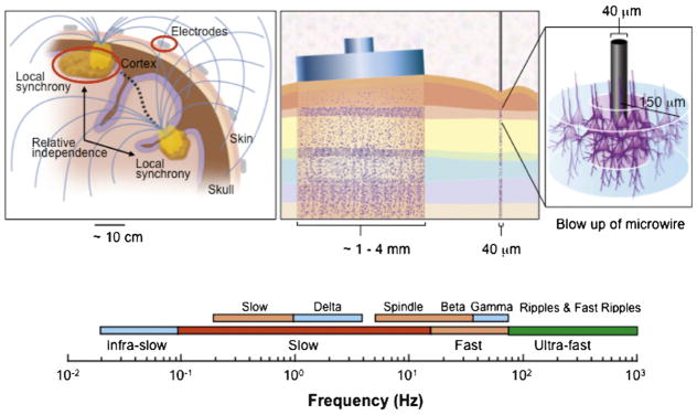
The spatial and temporal scales of human brain electrophysiology. Left: Figure indicating the ~7 cm2 patch of synchronous cortical activity required to generate a scalp detected EEG oscillation. Synchrony between distant regions of the brain are thought to support binding neuronal activity. Middle: The relative scale recorded from clinical macroelectrodes and subpial microwires. Right: Blow-up of penetrating microelectrode sampling activity from neurons within a radius of ~150 μm. Bottom: The wide range of spectral activity recorded from human brain from infra-slow oscillations to ultra-fast.