Fig. 8.
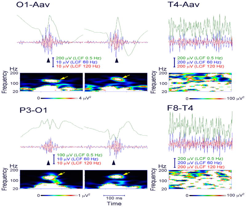
Ripple oscillations in the scalp EEG recorded from a child with Landau–Kleffner syndrome. Representative spikes (left and middle columns, arrowhead) are associated with ripple oscillation, which was largely invariant irrespective of low-cut frequency (LCF) of whether 60 or 120 Hz (EEG traces filtered at 0.5, 60, and 120 Hz are shown in green, blue, and red, respectively). The EEG was recorded during non-REM sleep and therefore did not include muscle activity or eye movements. Identical EEG data are presented in a referential montage (top: O1 with reference to the average EEG of bilateral earlobes, indicated as O1-Aav) and a bipolar montage (bottom: P3-O1). Note that spike-related ripples with at least four consecutive oscillations are clearly observed in both montages. Each panel of time–frequency spectra shows a corresponding discrete blob (arrow) with a frequency at around 130 Hz irrespective of referential or bipolar montage. In contrast, muscle activity (right column) contaminated to scalp EEG recorded during wakefulness is dominant over the temporal region (T4, F8-T4) close to muscles and has very irregular morphology and a noisy spectral pattern with no outstanding blobs.

