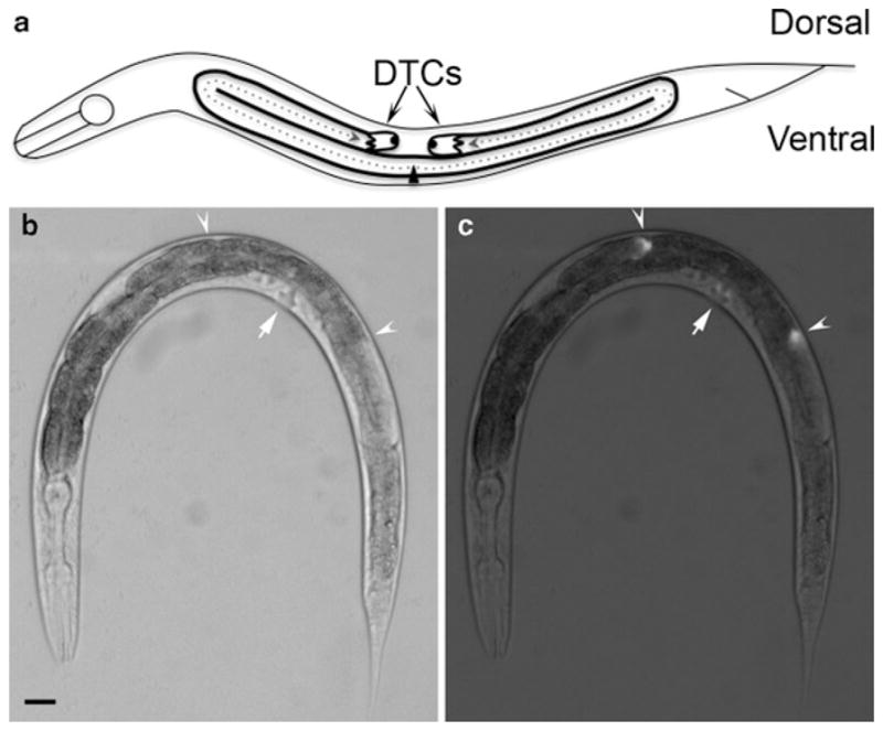
Fig. 1.

Distal tip cell migration and gonad morphology in the Caenorhabditis elegans hermaphrodite. (a) Schematic diagram of a late L4 animal. At the L2 larval stage, DTCs initiate migration on the ventral side (at the triangle, which represents the vulva) and follow the paths indicated by the dotted arrows. Migration terminates on the dorsal side opposite the vulva, yielding two U-shaped gonad arms. DIC image (b) and fluorescence superimposed on DIC image (c) of an L4 nematode carrying the lag-2p::GFP transgene. The lag-2 promoter is active in DTCs and drives the expression of GFP in these cells. Arrowheads indicate the DTCs and arrows indicate the location of the vulva. Anterior is to the left and ventral is down in all images. Scale bar represents 20 μm.
