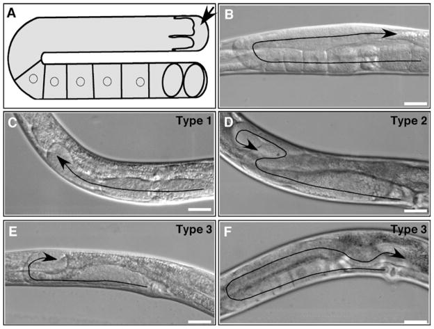
Fig. 4.
Examples of wild-type and defective DTC migration paths and gonad morphologies. (a) Diagram showing one arm of the hermaphrodite gonad. An arrow indicates the DTC. Cuboidal cells are maturing oocytes. Ovals are fertilized embryos. (b–f) DIC images showing wild-type morphology (b) and three types of defects (c–f). Treated rrf-3(pk1426) hermaphrodites were grown on Escherichia coli HT115(DE3) carrying an empty vector (b) or RNAi targeting vectors for act-1 (c), ced-5 (d), pat-3 (e), or dyn-1 (f). Arrows indicate DTC migration paths. Anterior is to the left, ventral is down in all images except (c) and (e) in which anterior is to the right. Bars, 20 μm. Adapted with permission from Journal of Cell Science (http://www.jcs.biologists.org) and originally published in Cram et al. (2006) (doi: 10.1242/jcs.03274) (25).

