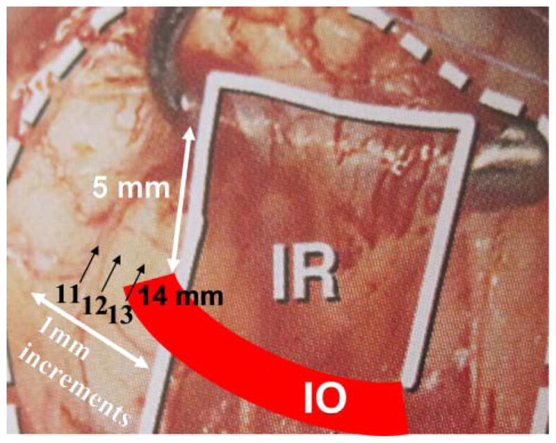
Figure 1.

Graded inferior oblique recession. Grading of inferior oblique recession by 1 mm increments from a 14 mm recession which is at the temporal inferior rectus (IR) border 5 mm posterior to the IR insertion. This figure is only reproduced in colour in the online version.
