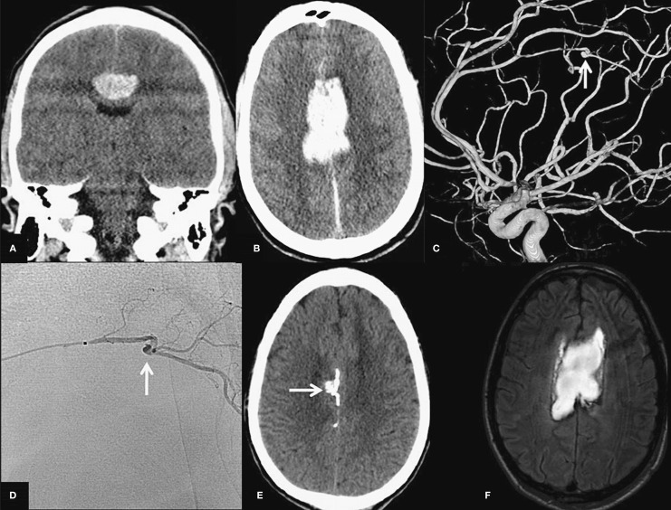
Figure 1.
Coronal (A) and transverse (B) CT scan with hematoma in the corpus callosum. C) Lateral view of right internal carotid 3D angiogram demonstrates a tiny pericallosal artery aneurysm (arrow). D) Lateral view of superselective angiogram shows the microcatheter positioned just proximal to the aneurysm. From this point glue was injected. The arrow points to the aneurysm. E) Native CT scan demonstrates radio-opaque glue in the aneurysm (arrow) and in proximal and distal parent vessels. F) T2-weighted MR after embolization shows hematoma with no infarction in distal anterior cerebral artery territory.

