
FIG. 1.
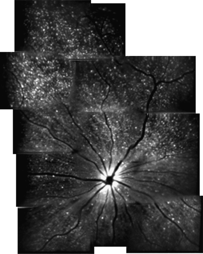
The detection of apoptosing retinal cells imaging approach for detecting retinal ganglion cell (RGC) apoptosis in animal models of ocular disease. Representative montage in vivo image of RGC apoptosis in a rat treated with intravitreally injected staurosporine. Imaging was accomplished using a confocal scanning laser ophthalmoscope with argon laser (488 nm) excitation of intravitreally injected Alexa Fluor 488-Annexin V conjugates, allowing visualization of apoptosing RGCs (hyperfluorescent white dots). Reproduced with permission from Cordeiro.24