
Fig. 2.
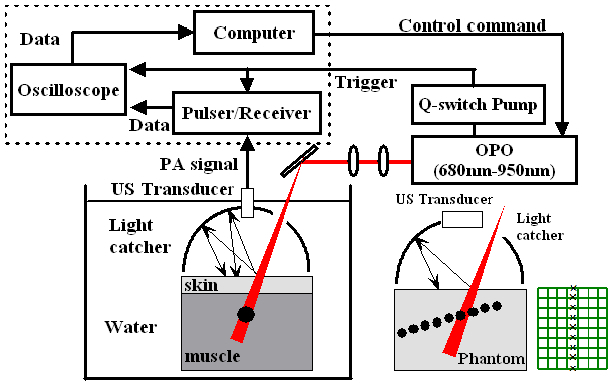
Experimental setup for in vitro photoacoustic imaging. (a) is the experimental setup using a single element transducer. (b) For 2D PA imaging, the dashed box in the block diagram was replaced with a commercial ultrasound scanner, a single element US transducer was replaced with a linear array transducer, and a black sheet of mesh was embedded in a gelatin phantom as a multiple point targets. Note a separate light catcher machined for the linear array transducer was used for 2D PA imaging.