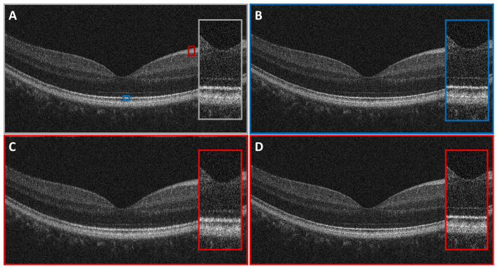
Fig. 6.
(A) An original OCT B-scan image of a normal human retina. Dispersion was approximately matched with a water cell in the OCT reference arm. (B) An OCT B-scan numerically dispersion compensated with the dispersion mismatch extracted from a single A-scan near the position indicated by the blue box in (A). Only the IS/OS was used for dispersion mismatch extraction. (C) An OCT B-scan numerically dispersion compensated with the dispersion mismatch extracted from a single A-scan near the position indicated by the red box in (A). The entire NFL was used for dispersion mismatch extraction. (D) The dispersion mismatch was extracted from 20 A-scans near the position indicated by the red box in (A) and averaged and filtered to reduce speckle.

