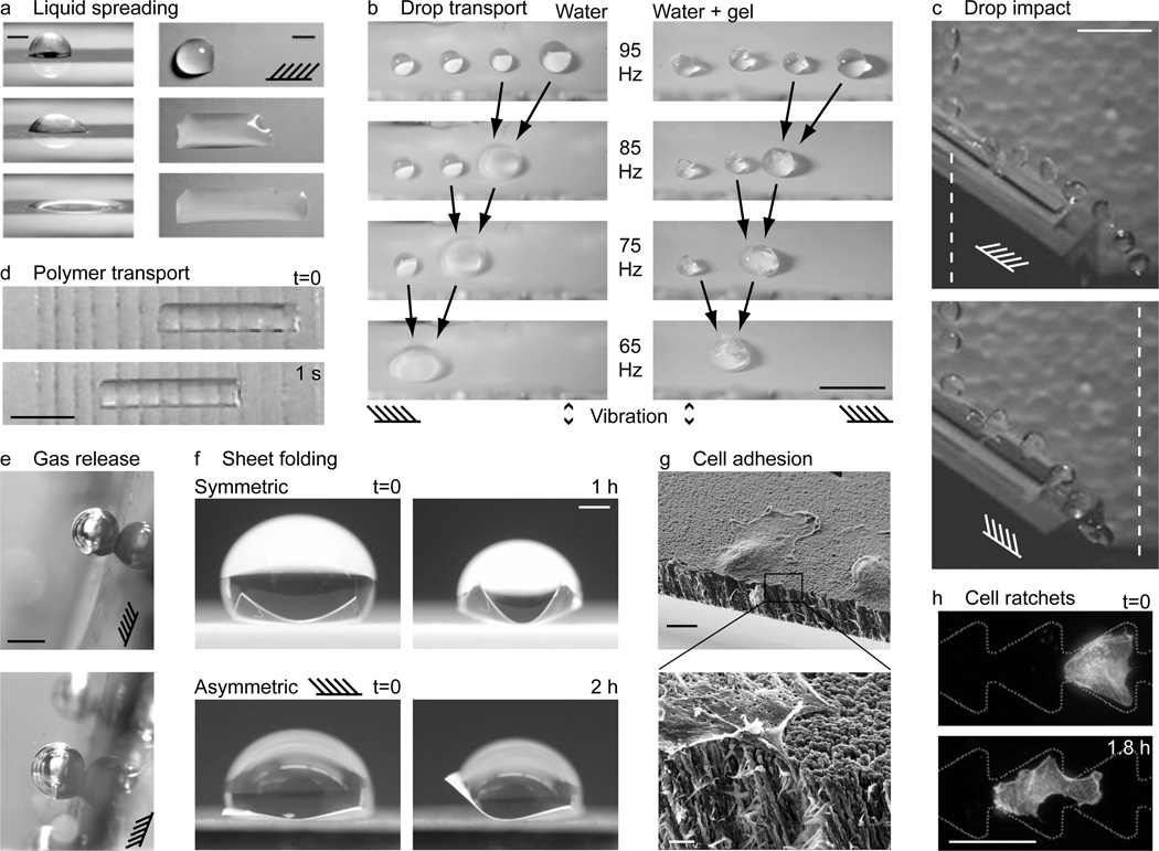
Figure 4.
Applications of directional surfaces. (a) Snapshots of a droplet spreading in one direction on a directional surface. Schematic indicates direction of nanorod tilt. Scale bars 1 mm. Source: Chu et al.,[42] reprinted with permission from Macmillan Publishers: Nature Materials, copyright 2010. (b) Drops and drops containing cargo being transported on directional surfaces forced with mechanical vibration. The forcing frequency was chosen close to the resonant frequency of the largest drop, and amplitude low enough so that smaller drops did not move. When two drops coalesced, their size increased and resonant frequency decreased. Thus, the forcing frequency was also reduced to keep the largest drop moving. Scale bar 5 mm. Reprinted with permission from Sekeroglu et al.[54] Copyright 2011, American Institute of Physics. (c) Drops impacting a directional surface with the grain (top) bounce further than those impacting against the grain (bottom). Dashed vertical lines demark the extent of the trajectory in the top image; schematics indicate the grain, i.e. nanorod direction. Scale bar 1 cm. Source: new unpublished work by the authors (see Experimental for details). (d) Soft gel transport on a PDMS surface with angled cuts aligned in the direction of motion. Snapshots shown 1 s apart. Scale bar 1 cm. Source: Mahadevan et al.,[55] copyright (2004) National Academy of Sciences, U.S.A. (e) Directional control of gas release. Gravity and buoyancy force point in (above) and against (below) the direction of nanorod tilt, as indicated by the schematic. Scale bar 1 mm. Source: new unpublished work by the authors (see Experimental for details). (f) Directional folding. Polymer sheets were folded by evaporating droplets. Folding was symmetric and asymmetric for sheets with isotropic and directional surfaces, respectively. Scale bar 4 mm. Source: new unpublished work by the authors (see Experimental for details). (g) Directional cell adhesion on a directional surface. Zoomed image indicates cell filopodia penetrating between the nanorods. Scale bars 10 µm (top) and 1 µm (bottom). Reprinted with permission from Christophis et al.[60] Copyright 2011, American Vacuum Society. (h) Channels with ratchet surfaces for cell transport. Scale bar 50 µm. Source: Mahmud et al.,[61] reprinted with permission from Macmillan Publishers: Nature Physics, copyright 2009.

