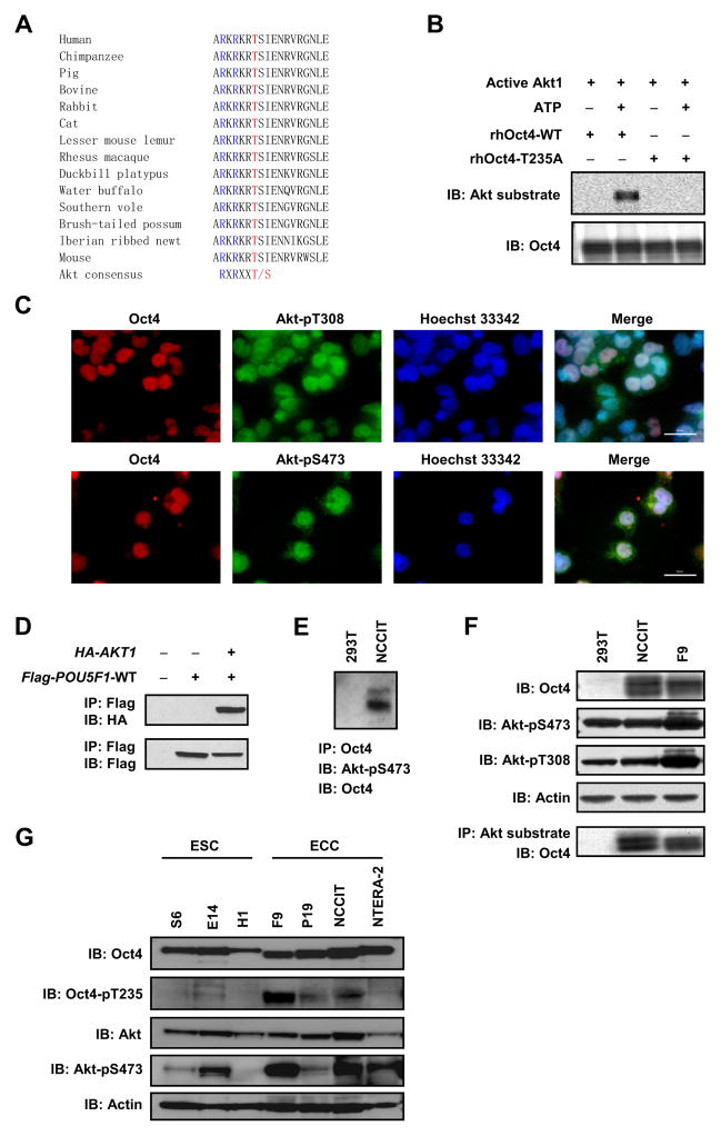
Figure 1. Akt phosphorylates Oct4 in ECCs.
(A) Amino acid sequence alignment of Oct4. (B) Purified recombinant human Oct4-WT (rhOct4-WT) or Oct4-T235A (rhOct4-T235A) protein was incubated with active Akt1 protein in the absence or presence of 200 μM ATP, and immunoblotted with the indicated antibodies. (C) NCCIT cells were co-immunostained with anti-Oct4/anti-Akt-pT308 (upper) or anti-Oct4/anti-Akt-pS473 (lower), and counterstained with Hoechst 33342. Bars, 50 μm. (D) Flag-POU5F1-WT and HA-AKT1 were co-transfected into 293T cells. Cell lysates were immunoprecipitated with anti-Flag M2 beads and immunoblotted with the indicated antibodies. (E) Cell lysates were immunoprecipitated with mouse anti-Oct4. Immuno-complexes were sequentially immunoblotted with rabbit anti-Akt-pS473 (upper band) and rabbit anti-Oct4 (lower band). (F) Whole cell lysates were immunoprecipitated with anti-phospho-Akt substrate rabbit mAb. Immuno-complexes were immunoblotted with mouse monoclonal anti-Oct4 (bottom panel). Upper panels are whole cell lysate immunoblots (IB) probed with the indicated antibodies. (G) Whole cell lysates were resolved by PAGE and immunoblotted with the indicated antibodies.

