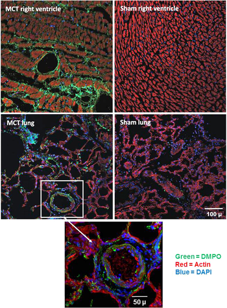
Fig. 5.
Monocrotaline-mediated free radical formation in rat lung and right ventricle. Three weeks after receiving monocrotaline (60 mg/kg ip) or saline (sham), rats received DMPO (2 g/kg total ip) in three doses 24, 12, and 6 h before sacrifice. At sacrifice, right ventricle (top) and lung tissue (bottom) were harvested, fixed, sectioned, and stained for DMPO–protein radicals using a primary rabbit polyclonal anti-DMPO Ab (green, DMPO; red, actin; and blue, DAPI nuclear stain). The white box and arrow indicate a magnified portion of the MCT-treated rat lung tissue. Data are representative images from three rats for MCT and three sham rats.

