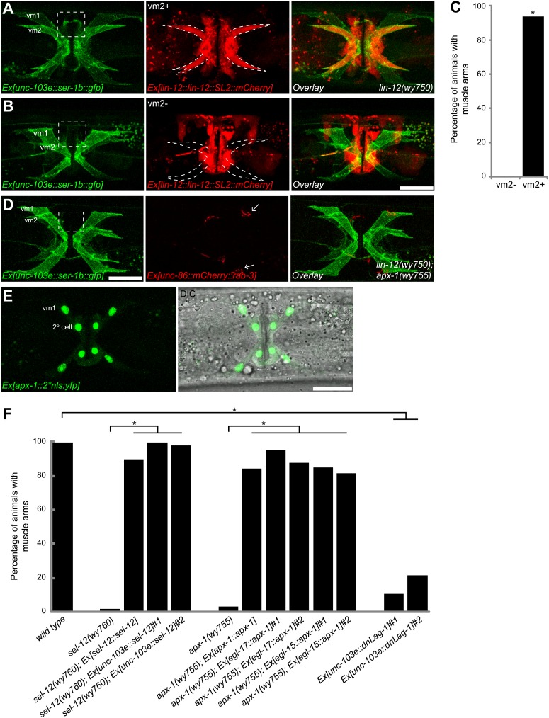
Figure 4. Cell-autonomous requirement of LIN-12 in vm2.
(A) and (B) Representative images of lin-12(wy750) mutant animals with non-mosaic (A) and mosaic (B) expression of lin-12::SL2::mCherry. Left column shows the vulval muscles labeled by SER-1B::GFP. Middle column shows the lin-12 expression patterns. Right column shows the overlaid images. Note that the complete lin-12 expression (vm2+) rescues muscle arm defects of lin-12(wy750) (A). Lacking of lin-12 expression in vm2 (vm2-) abolishes its capacity to rescue muscle arm defects (B). Boxed areas indicate synaptic regions. Dashed lines indicate the morphology of vm2. Scale bar is 20 μm. (C) Quantification of the vm2 muscle arm phenotypes in the mosaic transgenic animals. *p<0.0001, Fisher's exact test, n = 48–50. (D) Vulval muscle morphology in the apx-1(wy755); lin-12(w750) mutant animals. Boxed area indicates synaptic region. Arrows indicate HSN cell bodies. Scale bar is 20 μm. (E) Epifluorescence and DIC images showing the apx-1 expression pattern in young adult. The four cells in the center of the image are 2° vulval epithelial cells. The four cells at the periphery are vm1 cells. Scale bar is 20 μm. (F) Cell-autonomous requirement of apx-1, sel-12 and lag-1. Quantification of muscle arm phenotypes in animals with different genotypes indicated on the X-axis. *, P<0.0001, Fisher's exact test, n = 40–57.


