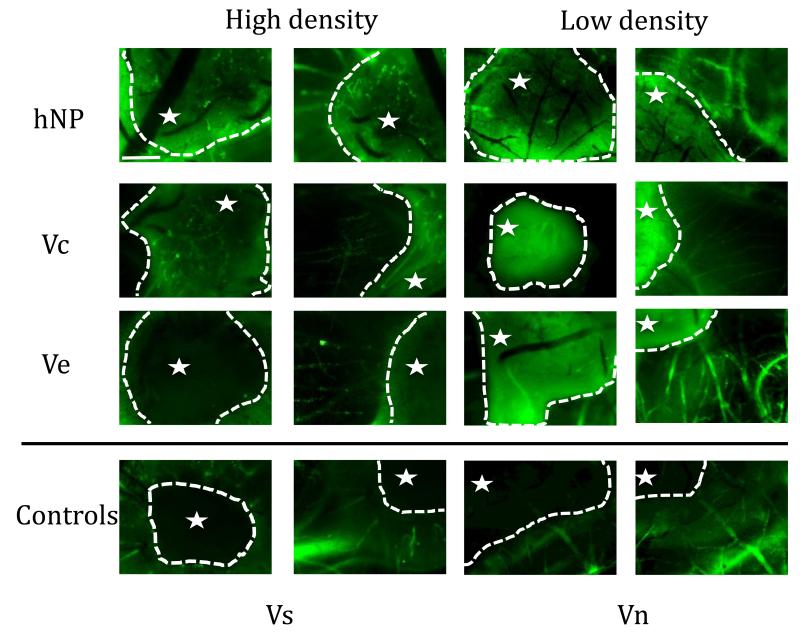
Figure 10.
CAM fluorescent micrographs of fibrin implant show that covalently bound VEGF leads to infiltration and branching of blood vessels within implant. Fluorescent micrographs were taken both of the implant itself and just outside the implant to show vessel morphology at the interface of the fibrin gel with the CAM (the star denotes the inside of the gel). Electrostatically bound VEGF releases and leads to radial vessel formation similar to the soluble VEGF condition (Scale bar = 25 μm).

