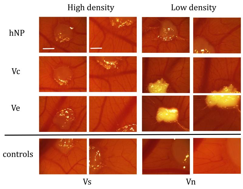
Figure 9.
CAM micrographs of fibrin implant and surrounding tissue show VEGF leads to induction of blood vessels surrounding implant. Since the low density conditions required more polystyrene particles, the fibrin gels became more opaque. The negative control, fibrin only (Vn), does not show the vascular induction of the other conditions with VEGF. Characteristic radial vessels originating from the implants are observed in every condition except the negative control (First and third column scale bar = 100 μm, second and fourth column scale bar = 60 μm).

