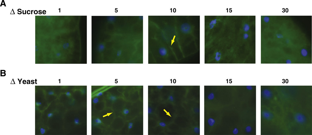
Fig. 4.
Insulin-signaling activity as a function of food content. Flies expressing the tGPH reporter construct were raised for ten days on ΔS diets (top panels) or ΔY diets (lower panels). Dissected fat bodies were evaluated for GFP staining in the membrane. Strongest membrane GFP staining is observed at intermediate food concentrations and declines both with low and high nutrient content (blue: DAPI; green: GFP; yellow arrows depict tGPH staining in the plasma membrane).

