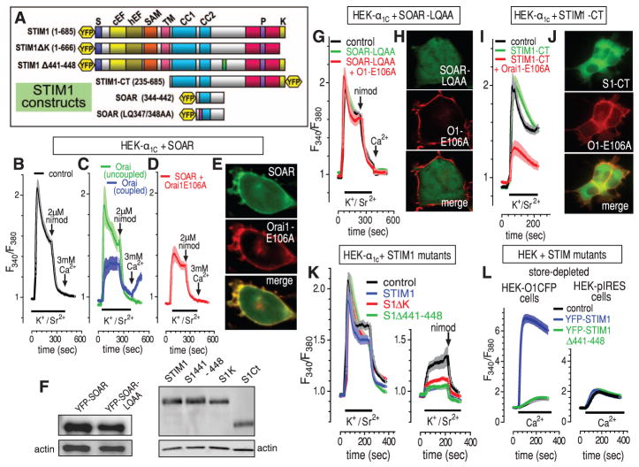
Fig. 4.
Defining the molecular domains of STIM1 mediating CaV1.2 channel inhibition. (A) STIM1 constructs used. (B to D) Fura-2 responses to K+/Sr2+ pulses (bars), 2 μM nimodipine, and 3 mM Ca2+ (arrows) in α1C-expressing HEK293 cells without cotransfection (B) or with cotransfection of YFP-SOAR (C) or YFP-SOAR + Orai1-E106A-CFP (D). Statistics shown in fig. S4A. In (C), Orai-coupled (n = 97; cells with Orai-mediated Ca2+ entry) and Orai-uncoupled (n = 135; cells with no Orai-mediated Ca2+ entry) are defined in the text. Their relative YFP-SOAR expression is shown in fig. S4B. (E) Localization of YFP-SOAR + Orai1-E106A-CFP cotransfected cells. (F) Western analysis of STIM construct expression. (G) Fura-2 responses in α1C-expressing HEK293 cells cotransfected with YFP-SOAR-LQ347/348AA alone or with Orai-E106A-CFP. (H) Imaging of the mutant SOAR + Orai–expressing cells in (G). (I) Fura-2 responses in α1C-expressing HEK293 cells cotransfected with YFP-STIM1-CT alone or with Orai-E106A-CFP. (J) Imaging of the STIM-CT + Orai–expressing cells in (I). (K) Fura-2 responses in α1C-expressing HEK293 cells before (left) and after (right) store depletion with 2 μM ionomycin; cells were cotransfected with either normal STIM1, the STIM1-ΔK truncation (Δ667-685), the STIM1-Δ441-448 deletion, or empty plasmid (statistics shown in fig. S4C). (L) Fura-2 responses to 1 mM Ca2+ addition for Orai1-CFP–expressing HEK293 cells (left) or control internal ribosomal entry site plasmid (pIRES)–HEK cells (right) transfected with YFP-STIM1-wild-type or YFP-STIM1-Δ441-448.

