

Abstract
Research using Xenopus takes advantage of large, abundant eggs, and readily manipulated embryos in addition to conserved cellular, developmental and genomic organization with mammals. Research on Xenopus has defined key principles of gene regulation and signal transduction, embryonic induction, morphogenesis and patterning as well as cell cycle regulation. Genomic and genetic advances in this system, including development of Xenopus tropicalis as a genetically tractable complement to the widely used Xenopus laevis, capitalize on the classical strengths and wealth of achievements. These attributes provide the tools to tackle the complex biological problems of the new century, including cellular reprogramming, organogenesis, regeneration, gene regulatory networks and protein interactions controlling growth and development, all of which provide insights into a multitude of human diseases and their potential treatments.
Basic features of Xenopus
‘I don’t see no p’ints about that frog that’s any better’n any other frog.’ So says the stranger before placing his wager in Mark Twain’s famous tale “The Celebrated Jumping Frog of Calaveras County” [1]. In modern biology there are practical advantages and limitations that dictate which animals are suited for particular problems, and new genomic and genetic tools have extended the historical advantages of amphibians. Xenopus has a leg up on other frogs, toads and salamanders, and offers particular experimental advantages over other vertebrate systems in general, centering on an abundance of large and robust eggs and embryos, accessible at all developmental stages. The conservation of key cellular and developmental processes and a high degree of genomic synteny with mammals provide powerful links for using Xenopus research to understand human development and disease.
Xenopus, like many amphibians, produces many (often thousands) of embryos that can be cultured in simple salt solutions, or eggs that can be crushed to make a versatile cell-free extract. In this review the genus Xenopus refers to work on two species, Xenopus laevis, the species first widely used by researchers, and Xenopus tropicalis, more recently adopted because of advantages for genetic and genomic work. Xenopus laevis first came to be widely used because it lays eggs year round in response to mammalian hormones, notably chorionic gonadotropin produced during pregnancy. The use of Xenopus laevis in human pregnancy testing also proved that this frog is tough and reliable, and therefore useful not only for clinical assays but also for research [2]. Three features of Xenopus eggs and embryos are especially important. Firstly, the embryos tolerate extensive surgical manipulations, ranging from very delicate procedures, such as transplantations of single cells, to extensive “cut and paste” operations that challenge large pieces of the embryo with new environments. Secondly, eggs and embryos are easily injected with material ranging from nuclei, as used in classic studies of animal cloning [3], to a variety of macromolecules, often nucleic acids and proteins, making this one of the best animal models for testing the functions of gene products. Targeting of injections to particular blastomeres, and therefore particular lineages, is also very useful (e.g. [4, 5]). Thirdly, eggs and embryos provide an abundant source of material for biochemical studies. For example, centrifuged eggs yield a cell-free extract that recapitulates in vitro the complex events of the cell cycle, and can be fractionated to identify structural and regulatory components [6]. The advantages extend to modern genomic analyses, e.g. performing analysis of gene expression and epigenetic changes in different dissected tissues (e.g. [7]).
Research using Xenopus has led to many breakthroughs in developmental and cell biology including fundamental discoveries regarding embryonic induction and patterning, signal transduction pathways controlling development, and the biochemistry of the cell cycle. Some of the major accomplishments are listed in Box 1.
Box 1. Major achievements of Xenopus research in cell and developmental biology, focusing on genetic and genomic approaches.
Xenopus research has played a prominent role in the recent history of cell and developmental biology. A partial list is shown below (citing only a few of the key papers). This list was generated in part from the 2011 Xenopus White Paper (www.xenbase.org), a community-generated document written to propose resources that will advance the NIH mission to understand living systems and apply new knowledge to improve human health.
| 1958 | Discovery that somatic nuclei, when transplanted into an egg, can fully reprogram development, demonstrating the principle of genomic equivalence of nuclei, and providing the basis for current work on animal cloning and nuclear reprogramming [8]. | |
| 1964–1968 | Discovery that the nucleolus is the site of the rRNA genes, and that these genes are amplified during oogenesis [9–11]. | |
| 1966 | Existence of mitochondrial DNA established and that it is maternally inherited [12]. | |
| 1968, 1971 | Isolation of the first eukaryotic genes (rRNA and 5SRNA genes) by equilibrium density centrifugation [13, 14]. | |
| 1971, 1977 | First eukaryotic translation and transcription-translation systems using the oocyte for injection and expression of mRNAs and cloned genes, respectively [15, 16]. | |
| 1976 | Discovery of MPF, a meiosis maturation promoting factor, a key insight for understanding mechanisms of cell cycle control [17]. | |
| 1977 | First system used for electrophysiological studies on cloned membrane channels and receptors[18]. | |
| 1978 | Identification of intrinsic nuclear targeting of nuclear proteins [19]. | |
| 1980 | Identification of the first eukaryotic transcription factor, TFIIIA [20]. | |
| 1983–1989 | First in vitro system for nuclear and chromatin assembly, and identification of key components of the cell cycle including its regulation by protein degradation of cyclins via ubiquitination [21, 22]. | |
| 1987 | Formation of mesoderm is mediated by members of the TGFβ and FGF growth factor families [23–25]. This work established the principle that peptide growth factors regulate many, if not most, tissue interactions controlling vertebrate embryo patterning and organogenesis. | |
| 1990’s | Identification of key genes involved in embryonic patterning, and development of the concept that many of these encode secreted growth factor antagonists (e.g. Noggin, and Cerberus, a potent head inducer [26]). | |
| 1996 | Identification of key genes that underlie establishment and patterning of the nervous system [27, 28]. | |
| 2005 | First genetic screen using wild-caught X. tropicalis [29]. | |
| 2006 | First genetic screen using ENU mutagenesis identifies 29 mutations in numerous organ systems [30]. | |
| 2009 | First mutant gene identified by positional cloning using new X. tropicalis genetic map [31, 32]. | |
| 2010 | X. tropicalis genome published, showing high conservation with mammalian genomes [33]. | |
Approaching genetic challenges in Xenopus laevis
Genetic research on Xenopus laevis is challenged by its allotetraploid genome (resulting from the hypothesized hybridization of two species [34]) yielding gene duplicates that would often preclude study of mutant phenotypes. In addition, X. laevis has a generation time of over a year. Nonetheless, some developmental mutations have been identified in X. laevis [35] and the anucleolate mutation, in which the nucleolus is lost, was particularly important in mapping ribosomal RNA genes to a unique locus, the “nucleolus organizer” [9].
Xenopus laevis has often been used for gain of function experiments, exploiting injection of mRNA into embryos to tease out mechanisms which control development. However, the complementary loss-of-function experiments were not readily addressed by mutagenesis until the introduction of Xenopus tropicalis as a model, (discussed in the next section). Manipulation of specific gene function was accomplished in Xenopus laevis by dominant negative constructs; these include the first such manipulation in vertebrates, demonstrating that FGF signaling is essential for the formation of most of the mesoderm [36]. The distinctions of TGFβ family signaling through Smad2 and Smad1 were also first clearly documented in Xenopus using a similar approach, and the resulting understanding of the roles of these signaling pathways to the organization of the embryo predated the loss-of-function experiments in other vertebrates [37].
Similar constructs revealed a novel role for the inhibition of BMP signaling in induction of the nervous system [38, 39], the first hint of what is now a mainstay in our understanding of signal transduction: that inhibition of signaling pathways is particularly important in embryonic patterning. This observation was significantly illuminated by the identification of a novel class of proteins whose sole function is to block such signaling. Among these is Noggin, a secreted protein that plays a key role in neural induction by blocking BMP signaling [40]. Noggin was identified by expression cloning [41], which has been used as a powerful genetic surrogate to identify novel genes and gene activities in many contexts. The approach entailed injection of pools of mRNAs produced from a gastrula cDNA library into embryos treated to block formation of the nervous system; pools of clones which rescued the phenotype were broken down to identify clones, and therefore genes, active in axis formation and neural induction. The method has been applied in numerous other contexts, for example, using the Xenopus laevis oocyte as an expression chamber to identify novel channels and transporters [42, 43].
EST projects and full-length cDNA sequencing efforts have enabled collections of full-length cDNA clones to be constructed ([44]; Xenopus Gene Collection: http://xgc.nci.nih.gov). The cDNAs, already in expression plasmids, provide easy access to most genes for expression as mRNAs, and a more systematic approach to expression cloning than was previously possible with random pools of cDNAs [45]. A recent extension of this work is the development of cDNA libraries containing all open reading frames (ORFs) in both X. laevis and X. tropicalis. The Xenopus ORFeome project will generate libraries flexibly designed to be used for a multitude of applications, including expression screening and proteomic screening, again ideally suited to the advantages of Xenopus.
In the absence of well-developed genetics, antisense oligonucleotides injected into eggs or embryos have become a mainstay in research on both Xenopus species. These are effective for the first few days of development (which includes the period of early organogenesis), such that antisense oligonucleotides have provided striking insights. An early example was the unexpected observation that an antisense oligonucleotide which destroyed maternal β-catenin mRNA was found to disturb axis formation, leading to the then novel insight that β-catenin plays a key role in Wnt signaling [46]. Blocking of β-catenin also was the basis for the first developmental application of morpholino oligonucleotides, which are effective post-fertilization, and have found wide application since [47]. While there are limitations in the effectiveness of these reagents, there are circumstances where their use goes beyond what could be normally expected of conventional genetics. For example, while the BMP signaling inhibitor, Noggin, is a potent neural-inducing molecule, Chordin and Follistatin also activate neural induction by blocking BMP signaling, and all three are implicated in the neural induction process. The functional redundancy of these proteins could be readily demonstrated by simultaneous antisense knockdown of noggin, chordin and follistatin, which blocks neural induction in vivo, a result that would be difficult to establish by crossing of genetic mutants [48].
Development of genetics in X. tropicalis
By the mid 1990’s, investigators seeking a genetically tractable amphibian species turned to Xenopus tropicalis, a west African relative of Xenopus laevis thought to have diverged approximately 50 mya [49], and which has a small, diploid genome [50] (Figure 1 shows both species). This rapidly developing animal circumvents the obstacles of the duplicated genome and longer generation time of X. laevis. X. tropicalis also enhances multigenerational studies in general, taking advantage of efficient transgenesis methods in Xenopus [51–54]. This has greatly enhanced the system for a multitude of assays, e.g. defining key regions in enhancers [55], production of transgenic reporter lines and development of methods for regulating gene expression (e.g. the Gal4-UAS system for conditional gene expression [56] and a number of Cre/loxP lines that can be used for fate mapping or targeting of gene constructs [57, 58]). While X. tropicalis eggs and embryos are smaller than X. laevis, they can nonetheless be effectively used both for embryological and biochemical work [59, 60].
Figure 1.
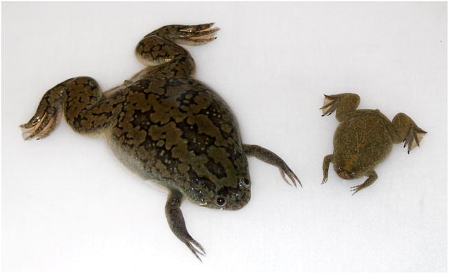
Photograph of adult X. laevis (left) together with X. tropicalis (right) (from ref. [61]). Development of both species is quite similar, though egg and embryo size is somewhat smaller for X. tropicalis and development can proceed more rapidly since the embryos are adapted to a higher temperature. Detailed features of the two systems have been described elsewhere [61].
Genetic and genomic approaches have been developed in parallel in order to jump-start this new system over the past decade. Beginning with the genetic side first, a number of strategies are being used to isolate or generate and screen recessive mutations in X. tropicalis. The availability of wild-caught animals initially presented a natural source of genetic variation to identify a group of recessive mutations in X. tropicalis [29, 62]. Such screens have been greatly facilitated by the use of gynogenetic diploid embryos (embryos that carry only maternal genes) by comparison with classical three generation screens to reveal recessive phenotypes in many organisms. This method was developed for Xenopus laevis in the 1970’s [63] and is accomplished by fertilizing eggs with UV-irradiated sperm, which activates development but does not contribute genetically to the embryo. While such embryos would normally be haploid, diploidy and normal development can be restored by preventing of release of the second polar body by applying high pressure or, more simply, by cold shock [64] to newly fertilized embryos. Segregation of maternal chromatid pairs reveals recessive mutations in the developing embryos. This method dramatically saves both time and space required by the conventional method of interbreeding offspring of heterozygous carrier animals. A wide range of developmental phenotypes have been reported, including defects in particular organ systems, like the inner ear, and very generalized defects, e.g. affecting left-right asymmetry [29]. An example of a mutation identified during a gynogenetic screen of non-inbred animals is cataract which shows opacity in the lens area resulting from a greatly reduced lens size. Whether this defect is autonomous to the lens, or due to failure in inductive signals from adjacent tissues, can be established because it is straightforward in Xenopus to make tissue chimeras (e.g. combining mutant lens tissue and wildtype inducers, or vice versa), as shown, for example, in other work relevant to eye development [65]. It is also valuable to note that, although Xenopus embryos are not transparent at pre-tadpole stages (as are zebrafish embryos), they become transparent as organogenesis proceeds and subtle morphological defects can be recognized.
In both Xenopus laevis and Xenopus tropicalis, the facility with which one can culture embryonic tissues, provides a means of scoring embryos for phenotypes (e.g. to look for defects associated with early morphogenesis). Combined with the relatively large size of cells, this has led to a much more detailed understanding of subcellular behaviors during morphogenesis than has been achieved in other vertebrates, for example in defining the role of planar cell polarity signaling in sustained and polarized cellular protrusions that direct gastrulation movements [66, 67].
More recent screens have taken advantage of inbred lines that have been developed in X. tropicalis, and using these for treatment with N-nitroso-N-ethylurea (ENU), the mutagen typically used in other vertebrates for generating high mutation rates. In the first published ENU screen, also a gynogenetic screen, 29 mutations were identified in a diversity of organ systems [30] (Figure 2). It should be noted that the long period of fertility for both X. laevis and X. tropicalis (ten years or more), greatly simplifies maintenance of stocks for backcrosses and test crosses, relative to other animal models.
Figure 2.

Summary of induced mutations found predominantly during the first ENU screen of X. tropicalis [30]. Mutations in a multitude of organ systems have been uncovered. Eight classes are presented here with images from one mutant in each class illustrated in this figure, and other members of the class denoted below or adjacent to the images. The images are paired: left is wildtype and right is mutant. Clockwise from upper left: eye phenotypes [brightfield of variegated retinal pigment epithelium in kaleidoscope (kal)]; inner ear [dysmorphic otoconia in komimi (kom)]; axial [the dwarf issunboushi (iss)]; neural crest [melanocytes in lumen of neural tube in cyd vicious (cyd)]; myofibrillogenesis [skeletal muscle stained with anti-alpha actinin (green) and phalloidin (red) showing disorganized sarcomeres in dicky ticker (dit)]; limb development [skeletal preparation showing complete absence of forelimb formation in xenopus de milo (xdm)]; cardiovascular [confocal image of muzak (muz) stained with anti-myosin heavy chain (green) and phalloidin (red)]; and blood [white hart (wha) showing reduced globin staining]. Figure courtesy of L. Zimmerman (National Institute for Medical Research, Mill Hill, London).
Genomic resources have been developed in parallel with genetic screening, and one area of recent and important confluence is the generation of a genetic map of simple sequence length polymorphisms (SSLP’s) [32], enabling positional cloning of a number of mutations. The initial stage of mapping to a chromosomal region is facilitated by the shortcuts associated with gynogenesis [68].
In the past two years the first positionally cloned genes have been identified [31, 69, 70]. While the phenotypes in screens of both wild-caught and ENU-treated animals have identified mutations that affect essentially every organ system, one mutation, xenopus de milo (xdm) (Figure 2), is of particular interest because it affects limb formation, highlighting that, among the non-mammalian genetic systems, uniquely valuable mutations will be forthcoming in the tetrapod Xenopus tropicalis. The accessibility of tissues in Xenopus embryos also provides advantages over other tetrapods for studying limb development. Analysis of limb development is also important as a basis for understanding limb regeneration, a fascinating feature of amphibians [71].
Because so many gene networks are already under study, in Xenopus and other model organisms, there is a pressing need for developing mutations in already identified genes. At present there are no homologous recombination strategies as in the mouse, where sophisticated gene modifications can be performed in embryonic stem cells. While embryonic stem cell equivalents have not been identified in Xenopus, genetically modified cultured cell nuclei can be reintroduced into eggs by nuclear transfer [72]. Using nuclei from appropriately mutated cells may allow researchers to exploit the amazing ability of Xenopus eggs to reprogram development under the direction of transplanted nuclei [73]. For now, like many model systems, Xenopus researchers are screening through DNAs from large populations of mutagenized animals by next-generation sequencing to identify mutations in important genes (often referred to as TILLING [74–76]). The efficient mutagenesis rate generated by ENU [30] and a highly efficient sperm-freezing method [77] enhance the utility of a sperm bank for long-term storage of samples for TILLING. The development of zinc-finger nucleases as a targeting system has also recently been shown to be applicable to X. tropicalis, where mutations in noggin have been induced [78]. The utility of an RNA interference strategy in Xenopus, and other vertebrates, is also of potentially high impact. Recent work using Xenopus [79, 80] offers hope that such a system might be optimized for targeting genes both early and late in development. These papers highlight the unique utility of Xenopus for ease of delivery of potential vectors, and the value of the extensive background that Xenopus DNA and RNA synthesis research provides for untangling nucleic acid processing mechanisms during development.
Classic strengths in a genomic context
The revolution in genomic and proteomic techniques has changed our vision of what is possible in any experimental system, and Xenopus is no exception, here adding new tools that build on already unique strengths of Xenopus, that together have the potential to transform vertebrate genome biology overall. The X. tropicalis genome sequence was published in 2010 [33], providing an essential tool for Xenopus researchers, but also revealing exciting information about genome organization. A striking observation is the extraordinary amount of synteny between frog and human genomes. Syntenic regions often span a hundred genes or more, with most of the large scaffolds (about half the genome) showing a high degree of colinearity. Around the centromere of human chromosome 1 the order of genes in approximately 150 Mb remains intact in X. tropicalis, despite the approximately 360 million years of evolution that separate these two species. With regard to gene and chromosome organization the Xenopus tropicalis genome represents a stunning model for evaluating human gene organization, taking advantage of both synteny with the human genome and the experimental manipulations, e.g. rapid transgenesis, possible in Xenopus. In addition this genome provides the raw material for evaluating the ancestral chromosome structure of tetrapods.
The evolutionary distance from mammals to frogs appears ideal for identification of conserved non-coding elements by bioinformatic comparisons. Studies of this kind reveal a high degree of conservation in putative regulatory elements surrounding developmental genes which might be expected to have conserved function, e.g. the Six3 gene [33], important for neural patterning in both frog and human, and the FoxE3 gene, essential for eye formation [55]. In other cases (e.g. the SCL locus, involved in blood, vasculature and brain development) there is conservation of function and expression but not as high a degree of sequence conservation in regulatory elements [81]. In all cases bioinformatic studies can be validated by transgenic assays, as discussed above. Because transgene integration occurs efficiently and early (at the one or two-cell stage), expression can be scored directly in the injected embryos (Figure 3). These approaches have been, and will be, invaluable in untangling the gene regulatory networks controlling early developmental decisions.
Figure 3.

Analysis of the Six3 locus in X. tropicalis (from ref. [33]). This analysis demonstrates that the X. tropicalis genome is well placed for comparative genomic studies to identify enhancers in conserved non-coding sequences (CNS) and that the fast transgenic methodology in Xenopus is particularly useful in testing their relevance as gene regulatory elements. In the middle of the figure one can see that in the 100 Kb surrounding the Six3 gene the mouse genome is so highly conserved compared to the base genome used here (human) that potentially functional conserved non-coding sequences (CNS) cannot be readily identified. However, when human is compared to Xenopus, seven highly conserved non-coding regions are found. Each was tested by co-transgenesis where a PCR product for the CNS is mixed with a basal promoter-green fluorescent protein (GFP) fusion construct in a suspension of sperm nuclei and injected into embryos, as summarized in the top part of the figure. Mixed enhancer and promoter fragments concatamerize (co-transgenesis) and are integrated rapidly, allowing efficient testing for enhancer activity in the conserved non-coding regions. At the top left, the endogenous expression of Six3 in eye and brain is shown by in situ hybridization. At the bottom of the figure, CNS3 and CNS5 were identified as enhancers of Six3 by the transgenic assay. The two enhancers together account for all embryonic expression of Six3. Note that the evolutionary distance of the Fugu genome is such that one of the key enhancers (location demarcated by red arrow) would not have been identified by this method.
X. tropicalis was just the first frog whose sequence was determined, and there used to be doubt that the allotetraploid genome of X. laevis could be properly assembled because of the confounding effect of very similar, though duplicated, genes. However, with the rapidly plummeting cost of DNA sequencing, and the appreciation that the duplicated alloalleles differ substantially in sequence [49] a high quality genome assembly will soon be available for X. laevis that will greatly enhance experiments in several ways. For example, it will be possible to design accurately morpholino oligonucleotides that target translation starts or splice junctions of both alloalleles, as well as to define a comprehensive proteome that will assist in assays where peptides are identified by mass spectrometry. Despite the possibility of setting up analogous cell free systems [82] or embryology in X. tropicalis [83], X. laevis embryos remain experimentally very attractive because they are larger, easier to manipulate, and also because they yield about five-fold more material per embryo, an asset for biochemical work. Furthermore, the cell free systems in X. tropicalis are not yet as reliable as those from X. laevis. Thus for biochemical and cell biological analysis, X. laevis will continue to be the preferred model system for proteome analysis, as it has already been for the cell cycle [84] or the analysis Wnt signaling dynamics [85]. Some issues, like scaling of organelle, cell or tissue size, for example determining what controls the different mitotic spindle size between the two species, highlight the value of comparative work using both species [60].
An area of intense interest in genome biology is the role of epigenetic changes in gene activity, especially in the context of development. Here, the genomic information from X. tropicalis is particularly revealing, through acquisition of a genome-wide gastrula stage transcriptome by RNA-seq in parallel with chromatin immunoprecipitation (ChIP- seq) to locate sites of histone modification [7]. Particular forms of modified histones are associated with localized gene expression, zygotic gene activation and regions of repression, revealing a global hierarchy in zygotic gene activation and localized expression. The ability to perform methods like ChIP-seq on dissected tissues highlights the value of the large, accessible and easily manipulated embryos in Xenopus tropicalis. Genome-wide analyses of DNA methylation complement this work, showing a surprisingly high degree of DNA methylation in early development, but a nonetheless relatively open, active state of chromatin at these stages, followed by re-establishment of methylation-dependent repression during the period of organogenesis. This pattern is conserved in human ES cells [86]. In this latter work, efficient transgenesis in Xenopus played a key role in testing hypotheses generated by bioinformatic data.
These analyses of epigenetic changes can be examined at a level that goes beyond other model organisms because of the rich history of nuclear transplantation studies to evaluate the developmental potential of nuclei from a wide array of developmental stages and tissues. In recent years, this classic work has been augmented by evaluating the changes in chromatin states bringing a profound new level of insight into nuclear reprogramming, demonstrating an epigenetic “memory” in transplanted nuclei not recognized earlier [87, 88]. These experiments are discussed in detail by Pasque et al. in this issue.
To complement the new techniques and information from genomics, a new U.S. National Xenopus Resource (NXR) at the Marine Biological Laboratory in Woods Hole will house genetically modified stocks and serve as a training resource and gathering place for disseminating new technology (http://www.mbl.edu/xenopus). The NXR will complement the European Xenopus Stock Center in Portsmouth, UK, providing new momentum for Xenopus researchers. In depth informatics resources developed in the community resource Xenbase (www.xenbase.org) also add an important dimension for researchers [89].
In a short review it is impossible to cover all aspects Xenopus research that exploit genetic and genomic tools but discussion of a few of these (Box 2) highlights some key areas where the new technologies will add great depth to the core strengths and intellectual base of this system.
Box 2. The impact of genetics and genomics on the spectrum of Xenopus research.
Modern Xenopus research spans a range of important biological problems, all of which are impacted by new genetic and genomic technologies. A few of these are described here briefly.
Mechanisms controlling the cell cycle and mitosis
A long-standing area of strength for the Xenopus system, new proteomic studies are already greatly enhancing our understanding of these problems (e.g [84]), and will be further enhanced by the X. laevis genome project.
Nuclear reprogramming and stem cell biology
Recent work clarifying epigenetic changes during nuclear reprogramming of somatic nuclei transplanted into the egg, will be greatly impacted by the genome-wide analyses (e.g. of histone methylation changes) now possible in Xenopus [7]. This combination of approaches, only feasible in Xenopus, has the potential to provide highly novel information for understanding stem cell biology in all animals. The unique accessibility of stem cells later in development as well, e.g. in the developing retina [90], offers unique prospects. As a complement to studies of mammalian stem cell mechanisms Xenopus is commonly used to test in vivo gene function because the frog embryo is so accessible and assayable throughout development [91].
Organogenesis
Access and the ease of transgenesis make Xenopus a powerful system for examining organogenesis, illustrated by recent work on pancreas formation [92] and ability to reprogram other endodermal derivatives to form pancreas [93]. The use of lineage-labeled transgenic lines in genetic screens will enhance identification of mutations in particular organ systems.
Tissue Remodeling during Metamorphosis and Regeneration
Xenopus metamorphosis is an accessible and easily manipulated system for studying tissue remodeling, where access to genomic tools is invaluable [94]. Amphibians are also unique among vertebrates for their regenerative ability, and recent work highlights novel insights into pathways regulating tissue regeneration [71, 95].
Xenopusas a model for study of human disease
Transgenes expressed in Xenopus have been found to mimic human genetic lesions (e.g. [96]), and gene products that regulate key developmental and cellular events, e.g. neural crest formation and cell movement and ciliogenesis, and are also involved in related human syndromes ([97] and [98], respectively). Mutations found in Xenopus genetic screens often appear to be linked to human syndromes as well [31].
Concluding remarks
As researchers delve into increasingly complex phenomena in cells and embryos it will become ever more important to examine these events in the living animal where the subtleties of these processes are revealed most clearly. This requires animals in which one can use every methodology possible for studying biological phenomena. Xenopus now stands out in this way, with the addition of genetic and genomic approaches that can build on the classical strengths and research contributions of this system. Genetic manipulation, coupled with the ability to take explants and carry out grafts, has led to enormous progress in teasing apart the signaling pathways that control early development. While the classical approaches of experimental embryology became unfashionable for a short while, every new technique that is applied to explants or cut and paste experiments illustrate the power of deconstructing the embryo in this manner. Explants allow high resolution imaging, and grafts or recombined explants permit a biochemical level of understanding of signaling and response between tissues. The activities and responses of transcription factors are now being assembled into gene regulatory networks [92]. This regulatory logic will be combined with the progress in cell biology to provide a satisfying understanding of the developmental program, and will provide a template for understanding the more complex events of commitment, morphogenesis, organogenesis, regeneration and human disease that still elude us and remain challenges for the future.
Acknowledgments
The authors would particularly like to acknowledge support of community resources by the National Institutes of Health and the genome sequencing by the Department of Energy’s Joint Genome Institute. The National Xenopus Resource and Development of a national TILLING resource are supported by grants RR025867 and HD065713, respectively, to RMG. A Xenopus tropicalis genome improvement grant and a Xenopus laevis genome sequencing project are supported by grants GM086321 and HD065705, respectively, to RMH and Daniel Rokhsar. The authors also gratefully research support, grants EY019000 and EY017400 to RMG and grants DC010210, GM042341, and GM049346 to RMH.
Footnotes
Publisher's Disclaimer: This is a PDF file of an unedited manuscript that has been accepted for publication. As a service to our customers we are providing this early version of the manuscript. The manuscript will undergo copyediting, typesetting, and review of the resulting proof before it is published in its final citable form. Please note that during the production process errors may be discovered which could affect the content, and all legal disclaimers that apply to the journal pertain.
References
- 1.Twain M. The Celebrated Jumping Frog of Calaveras County: And Other Sketches. C.H. Webb; 1867. [Google Scholar]
- 2.Gurdon JB, Hopwood N. The introduction of Xenopus laevis into developmental biology: of empire, pregnancy testing and ribosomal genes. Int J Dev Biol. 2000;44:43–50. [PubMed] [Google Scholar]
- 3.Gurdon JB, Uehlinger V. “Fertile” intestine nuclei. Nature. 1966;210:1240–1241. doi: 10.1038/2101240a0. [DOI] [PubMed] [Google Scholar]
- 4.Moody SA. Fates of the blastomeres of the 32-cell-stage Xenopus embryo. Dev Biol. 1987;122:300–319. doi: 10.1016/0012-1606(87)90296-x. [DOI] [PubMed] [Google Scholar]
- 5.Dale L, Slack JM. Fate map for the 32-cell stage of Xenopus laevis. Development. 1987;99:527–551. doi: 10.1242/dev.99.4.527. [DOI] [PubMed] [Google Scholar]
- 6.Philpott A, Yew PR. The Xenopus cell cycle: an overview. Methods Mol Biol. 2005;296:95–112. doi: 10.1385/1-59259-857-9:095. [DOI] [PubMed] [Google Scholar]
- 7.Akkers RC, et al. A hierarchy of H3K4me3 and H3K27me3 acquisition in spatial gene regulation in Xenopus embryos. Dev Cell. 2009;17:425–434. doi: 10.1016/j.devcel.2009.08.005. [DOI] [PMC free article] [PubMed] [Google Scholar]
- 8.Gurdon JB, et al. Sexually mature individuals of Xenopus laevis from the transplantation of single somatic nuclei. Nature. 1958;182:64–65. doi: 10.1038/182064a0. [DOI] [PubMed] [Google Scholar]
- 9.Brown DD, Gurdon JB. Absence Of Ribosomal Rna Synthesis In The Anucleolate Mutant Of Xenopus Laevis. Proc Natl Acad Sci U S A. 1964;51:139–146. doi: 10.1073/pnas.51.1.139. [DOI] [PMC free article] [PubMed] [Google Scholar]
- 10.Brown DD, Dawid IB. Specific gene amplification in oocytes. Oocyte nuclei contain extrachromosomal replicas of the genes for ribosomal RNA. Science. 1968;160:272–280. doi: 10.1126/science.160.3825.272. [DOI] [PubMed] [Google Scholar]
- 11.Gall JG. Differential synthesis of the genes for ribosomal RNA during amphibian oogenesis. Proc Natl Acad Sci U S A. 1968;60:553–560. doi: 10.1073/pnas.60.2.553. [DOI] [PMC free article] [PubMed] [Google Scholar]
- 12.Dawid IB. Evidence for the mitochondrial origin of frog egg cytoplasmic DNA. Proc Natl Acad Sci U S A. 1966;56:269–276. doi: 10.1073/pnas.56.1.269. [DOI] [PMC free article] [PubMed] [Google Scholar]
- 13.Birnstiel M, et al. Properties and composition of the isolated ribosomal DNA satellite of Xenopus laevis. Nature. 1968;219:454–463. doi: 10.1038/219454a0. [DOI] [PubMed] [Google Scholar]
- 14.Brown DD, et al. Purification and some characteristics of 5S DNA from Xenopus laevis. Proc Natl Acad Sci U S A. 1971;68:3175–3179. doi: 10.1073/pnas.68.12.3175. [DOI] [PMC free article] [PubMed] [Google Scholar]
- 15.Gurdon JB, et al. Use of frog eggs and oocytes for the study of messenger RNA and its translation in living cells. Nature. 1971;233:177–182. doi: 10.1038/233177a0. [DOI] [PubMed] [Google Scholar]
- 16.De Robertis EM, Mertz JE. Coupled transcription-translation of DNA injected into Xenopus oocytes. Cell. 1977;12:175–182. doi: 10.1016/0092-8674(77)90195-7. [DOI] [PubMed] [Google Scholar]
- 17.Wasserman WJ, Masui Y. A cytoplasmic factor promoting oocyte maturation: its extraction and preliminary characterization. Science. 1976;191:1266–1268. doi: 10.1126/science.1083070. [DOI] [PubMed] [Google Scholar]
- 18.Kusano K, et al. Acetylcholine receptors in the oocyte membrane. Nature. 1977;270:739–741. doi: 10.1038/270739a0. [DOI] [PubMed] [Google Scholar]
- 19.De Robertis EM, et al. Intracellular migration of nuclear proteins in Xenopus oocytes. Nature. 1978;272:254–256. doi: 10.1038/272254a0. [DOI] [PubMed] [Google Scholar]
- 20.Engelke DR, et al. Specific interaction of a purified transcription factor with an internal control region of 5S RNA genes. Cell. 1980;19:717–728. doi: 10.1016/s0092-8674(80)80048-1. [DOI] [PubMed] [Google Scholar]
- 21.Lohka MJ, Masui Y. Formation in vitro of sperm pronuclei and mitotic chromosomes induced by amphibian ooplasmic components. Science. 1983;220:719–721. doi: 10.1126/science.6601299. [DOI] [PubMed] [Google Scholar]
- 22.Murray AW, Kirschner MW. Cyclin synthesis drives the early embryonic cell cycle. Nature. 1989;339:275–280. doi: 10.1038/339275a0. [DOI] [PubMed] [Google Scholar]
- 23.Smith JC. A mesoderm inducing factor is produced by a Xenopus cell line. Development. 1987;99:3–14. doi: 10.1242/dev.99.1.3. [DOI] [PubMed] [Google Scholar]
- 24.Slack JM, et al. Mesoderm induction in early Xenopus embryos by heparin-binding growth factors. Nature. 1987;326:197–200. doi: 10.1038/326197a0. [DOI] [PubMed] [Google Scholar]
- 25.Kimelman D, Bjornson C. Vertebrate mesoderm induction: from frogs to mice. Cold Spring Harbor Laboratory Press; 2004. [Google Scholar]
- 26.Bouwmeester T, et al. Cerberus is a head-inducing secreted factor expressed in the anterior endoderm of Spemann’s organizer. Nature. 1996;382:595–601. doi: 10.1038/382595a0. [DOI] [PubMed] [Google Scholar]
- 27.Zimmerman LB, et al. The Spemann organizer signal noggin binds and inactivates bone morphogenetic protein 4. Cell. 1996;86:599–606. doi: 10.1016/s0092-8674(00)80133-6. [DOI] [PubMed] [Google Scholar]
- 28.Piccolo S, et al. Dorsoventral patterning in Xenopus: inhibition of ventral signals by direct binding of chordin to BMP-4. Cell. 1996;86:589–598. doi: 10.1016/s0092-8674(00)80132-4. [DOI] [PMC free article] [PubMed] [Google Scholar]
- 29.Noramly S, et al. A gynogenetic screen to isolate naturally occurring recessive mutations in Xenopus tropicalis. Mech Dev. 2005;122:273–287. doi: 10.1016/j.mod.2004.11.001. [DOI] [PubMed] [Google Scholar]
- 30.Goda T, et al. Genetic screens for mutations affecting development of Xenopus tropicalis. PLoS Genet. 2006;2:e91. doi: 10.1371/journal.pgen.0020091. [DOI] [PMC free article] [PubMed] [Google Scholar]
- 31.Abu-Daya A, et al. Absence of heartbeat in the Xenopus tropicalis mutation muzak is caused by a nonsense mutation in cardiac myosin myh6. Dev Biol. 2009;336:20–29. doi: 10.1016/j.ydbio.2009.09.019. [DOI] [PMC free article] [PubMed] [Google Scholar]
- 32.Wells DE, et al. A genetic map of Xenopus tropicalis. Dev Biol. 2011;354:1–8. doi: 10.1016/j.ydbio.2011.03.022. [DOI] [PMC free article] [PubMed] [Google Scholar]
- 33.Hellsten U, et al. The genome of the Western clawed frog Xenopus tropicalis. Science. 2010;328:633–636. doi: 10.1126/science.1183670. [DOI] [PMC free article] [PubMed] [Google Scholar]
- 34.Kobel HR. Allopolyploid Speciation. In: Tinsley RC, Kobel HR, editors. The Biology of Xenopus. Clarendon Press; 1996. pp. 391–402. [Google Scholar]
- 35.Krotoski DM, et al. Developmental mutants isolated from wild-caught Xenopus laevis by gynogenesis and inbreeding. Journal of Experimental Zoology. 1985;233:443–449. doi: 10.1002/jez.1402330313. [DOI] [PubMed] [Google Scholar]
- 36.Amaya E, et al. Expression of a dominant negative mutant of the FGF receptor disrupts mesoderm formation in Xenopus embryos. Cell. 1991;66:257–270. doi: 10.1016/0092-8674(91)90616-7. [DOI] [PubMed] [Google Scholar]
- 37.Whitman M. Smads and early developmental signaling by the TGFbeta superfamily. Genes Dev. 1998;12:2445–2462. doi: 10.1101/gad.12.16.2445. [DOI] [PubMed] [Google Scholar]
- 38.Wilson PA, Hemmati-Brivanlou A. Induction of epidermis and inhibition of neural fate by Bmp-4. Nature. 1995;376:331–333. doi: 10.1038/376331a0. [DOI] [PubMed] [Google Scholar]
- 39.Sasai Y, et al. Regulation of neural induction by the Chd and Bmp-4 antagonistic patterning signals in Xenopus. Nature. 1995;377:757. doi: 10.1038/377757a0. [DOI] [PubMed] [Google Scholar]
- 40.Lamb TM, et al. Neural induction by the secreted polypeptide noggin. Science. 1993;262:713–718. doi: 10.1126/science.8235591. [DOI] [PubMed] [Google Scholar]
- 41.Smith WC, Harland RM. Expression cloning of noggin, a new dorsalizing factor localized to the Spemann organizer in Xenopus embryos. Cell. 1992;70:829–840. doi: 10.1016/0092-8674(92)90316-5. [DOI] [PubMed] [Google Scholar]
- 42.Boulter J, Boyer C. Expression cloning of neural genes using Xenopus laevis oocytes. Curr Protoc Neurosci. 2001;Chapter 4(Unit 4):3. doi: 10.1002/0471142301.NS0403s00. [DOI] [PubMed] [Google Scholar]
- 43.Bianchi L, Driscoll M. Heterologous expression of C. elegans ion channels in Xenopus oocytes. WormBook; 2006. pp. 1–16. [DOI] [PMC free article] [PubMed] [Google Scholar]
- 44.Gilchrist MJ, et al. Defining a large set of full-length clones from a Xenopus tropicalis EST project. Dev Biol. 2004;271:498–516. doi: 10.1016/j.ydbio.2004.04.023. [DOI] [PubMed] [Google Scholar]
- 45.Chen JA, et al. Identification of novel genes affecting mesoderm formation and morphogenesis through an enhanced large scale functional screen in Xenopus. Mech Dev. 2005;122:307–331. doi: 10.1016/j.mod.2004.11.008. [DOI] [PubMed] [Google Scholar]
- 46.Heasman J, et al. Overexpression of cadherins and underexpression of beta-catenin inhibit dorsal mesoderm induction in early Xenopus embryos. Cell. 1994;79:791–803. doi: 10.1016/0092-8674(94)90069-8. [DOI] [PubMed] [Google Scholar]
- 47.Heasman J. Morpholino oligos: making sense of antisense? Dev Biol. 2002;243:209–214. doi: 10.1006/dbio.2001.0565. [DOI] [PubMed] [Google Scholar]
- 48.Khokha MK, et al. Depletion of three BMP antagonists from Spemann’s organizer leads to a catastrophic loss of dorsal structures. Dev Cell. 2005;8:401–411. doi: 10.1016/j.devcel.2005.01.013. [DOI] [PubMed] [Google Scholar]
- 49.Hellsten U, et al. Accelerated gene evolution and subfunctionalization in the pseudotetraploid frog Xenopus laevis. BMC biology. 2007;5:31. doi: 10.1186/1741-7007-5-31. [DOI] [PMC free article] [PubMed] [Google Scholar]
- 50.Amaya E, et al. Frog genetics: Xenopus tropicalis jumps into the future. Trends in Genetics. 1998;14:253–255. doi: 10.1016/s0168-9525(98)01506-6. [DOI] [PubMed] [Google Scholar]
- 51.Kroll KL, Amaya E. Transgenic Xenopus embryos from sperm nuclear transplantations reveal FGF signaling requirements during gastrulation. Development. 1996;122:3173–3183. doi: 10.1242/dev.122.10.3173. [DOI] [PubMed] [Google Scholar]
- 52.Ogino H, et al. High-throughput transgenesis in Xenopus using I-SceI meganuclease. Nat Protoc. 2006;1:1703–1710. doi: 10.1038/nprot.2006.208. [DOI] [PubMed] [Google Scholar]
- 53.Pan FC, et al. I-SceI meganuclease-mediated transgenesis in Xenopus. Dev Dyn. 2006;235:247–252. doi: 10.1002/dvdy.20608. [DOI] [PubMed] [Google Scholar]
- 54.Hamlet MR, et al. Tol2 transposon-mediated transgenesis in Xenopus tropicalis. Genesis. 2006;44:438–445. doi: 10.1002/dvg.20234. [DOI] [PubMed] [Google Scholar]
- 55.Ogino H, et al. Convergence of a head-field selector Otx2 and Notch signaling: a mechanism for lens specification. Development. 2008;135:249–258. doi: 10.1242/dev.009548. [DOI] [PMC free article] [PubMed] [Google Scholar]
- 56.Chae J, et al. Inducible control of tissue-specific transgene expression in Xenopus tropicalis transgenic lines. Mech Dev. 2002;117:235–241. doi: 10.1016/s0925-4773(02)00219-8. [DOI] [PubMed] [Google Scholar]
- 57.Roose M, et al. Heat-shock inducible Cre strains to study organogenesis in transgenic Xenopus laevis. Transgenic research. 2009;18:595–605. doi: 10.1007/s11248-009-9253-4. [DOI] [PubMed] [Google Scholar]
- 58.Rankin SA, et al. Improved cre reporter transgenic Xenopus. Dev Dyn. 2009;238:2401–2408. doi: 10.1002/dvdy.22043. [DOI] [PMC free article] [PubMed] [Google Scholar]
- 59.Offield MF, et al. The development of Xenopus tropicalis transgenic lines and their use in studying lens developmental timing in living embryos. Development. 2000;127:1789–1797. doi: 10.1242/dev.127.9.1789. [DOI] [PubMed] [Google Scholar]
- 60.Levy DL, Heald R. Nuclear size is regulated by importin alpha and Ntf2 in Xenopus. Cell. 2010;143:288–298. doi: 10.1016/j.cell.2010.09.012. [DOI] [PMC free article] [PubMed] [Google Scholar]
- 61.Hirsch N, et al. Xenopus, the next generation: X. tropicalis genetics and genomics. Dev Dyn. 2002;225:422–433. doi: 10.1002/dvdy.10178. [DOI] [PubMed] [Google Scholar]
- 62.Grammer TC, et al. Identification of mutants in inbred Xenopus tropicalis. Mech Dev. 2005;122:263–272. doi: 10.1016/j.mod.2004.11.003. [DOI] [PubMed] [Google Scholar]
- 63.Tompkins R. Triploid and gynogenetic diploid Xenopus laevis. Journal of Experimental Zoology. 1978;203:251–256. [Google Scholar]
- 64.Kawahara H. Production of triploid and gynogenetic diploid Xenopus by cold treatment. Develop, Growth and Differ. 1978;20:227–236. doi: 10.1111/j.1440-169X.1978.00227.x. [DOI] [PubMed] [Google Scholar]
- 65.Henry JJ, Grainger RM. Early tissue interactions leading to embryonic lens formation in Xenopus laevis. Developmental Biology. 1990;141:149–163. doi: 10.1016/0012-1606(90)90110-5. [DOI] [PubMed] [Google Scholar]
- 66.Wallingford JB, et al. Dishevelled controls cell polarity during Xenopus gastrulation. Nature. 2000;405:81–85. doi: 10.1038/35011077. [DOI] [PubMed] [Google Scholar]
- 67.Goto T, et al. Planar cell polarity genes regulate polarized extracellular matrix deposition during frog gastrulation. Curr Biol. 2005;15:787–793. doi: 10.1016/j.cub.2005.03.040. [DOI] [PubMed] [Google Scholar]
- 68.Khokha MK, et al. Rapid gynogenetic mapping of Xenopus tropicalis mutations to chromosomes. Dev Dyn. 2009;238:1398–1346. doi: 10.1002/dvdy.21965. [DOI] [PMC free article] [PubMed] [Google Scholar]
- 69.Abu-Daya A, et al. The secreted integrin ligand nephronectin is necessary for forelimb formation in Xenopus tropicalis. Dev Biol. 2011;349:204–212. doi: 10.1016/j.ydbio.2010.10.015. [DOI] [PMC free article] [PubMed] [Google Scholar]
- 70.Geach TJ, Zimmerman LB. Paralysis and delayed Z-disc formation in the Xenopus tropicalis unc45b mutant dicky ticker. BMC Dev Biol. 2010;10:75. doi: 10.1186/1471-213X-10-75. [DOI] [PMC free article] [PubMed] [Google Scholar]
- 71.Beck CW, et al. Beyond early development: Xenopus as an emerging model for the study of regenerative mechanisms. Dev Dyn. 2009;238:1226–1248. doi: 10.1002/dvdy.21890. [DOI] [PubMed] [Google Scholar]
- 72.Kroll KL, Gerhart JC. Transgenic X. laevis embryos from eggs transplanted with nuclei of transfected cultured cells. Science. 1994;266:650–653. doi: 10.1126/science.7939720. [DOI] [PubMed] [Google Scholar]
- 73.Gurdon JB, et al. The developmental capacity of nuclei transplanted from keratinized skin cells of adult frogs. J Embryol Exp Morphol. 1975;34:93–112. [PubMed] [Google Scholar]
- 74.Stemple DL. TILLING--a high-throughput harvest for functional genomics. Nat Rev Genet. 2004;5:145–150. doi: 10.1038/nrg1273. [DOI] [PubMed] [Google Scholar]
- 75.Moens CB, et al. Reverse genetics in zebrafish by TILLING. Brief Funct Genomic Proteomic. 2008;7:454–459. doi: 10.1093/bfgp/eln046. [DOI] [PMC free article] [PubMed] [Google Scholar]
- 76.Comai L, Henikoff S. TILLING: practical single-nucleotide mutation discovery. Plant J. 2006;45:684–694. doi: 10.1111/j.1365-313X.2006.02670.x. [DOI] [PubMed] [Google Scholar]
- 77.Sargent MG, Mohun TJ. Cryopreservation of sperm of Xenopus laevis and Xenopus tropicalis. Genesis. 2005;41:41–46. doi: 10.1002/gene.20092. [DOI] [PubMed] [Google Scholar]
- 78.Young JJ, et al. Efficient targeted gene disruption in the soma and germ line of the frog Xenopus tropicalis using engineered zinc-finger nucleases. Proc Natl Acad Sci U S A. 2011;108:7052–7057. doi: 10.1073/pnas.1102030108. [DOI] [PMC free article] [PubMed] [Google Scholar]
- 79.Lund E, et al. Limiting Ago protein restricts RNAi and microRNA biogenesis during early development in Xenopus laevis. Genes Dev. 2011;25:1121–1131. doi: 10.1101/gad.2038811. [DOI] [PMC free article] [PubMed] [Google Scholar]
- 80.Chen CM, et al. Co-expression of Argonaute2 Enhances Short Hairpin RNA-induced RNA Interference in Xenopus CNS Neurons In Vivo. Frontiers in neuroscience. 2009;3:63. doi: 10.3389/neuro.17.001.2009. [DOI] [PMC free article] [PubMed] [Google Scholar]
- 81.Gottgens B, et al. Analysis of vertebrate SCL loci identifies conserved enhancers. Nat Biotechnol. 2000;18:181–186. doi: 10.1038/72635. [DOI] [PubMed] [Google Scholar]
- 82.Brown KS, et al. Xenopus tropicalis egg extracts provide insight into scaling of the mitotic spindle. J Cell Biol. 2007;176:765–770. doi: 10.1083/jcb.200610043. [DOI] [PMC free article] [PubMed] [Google Scholar]
- 83.Hirsch N, et al. Xenopus tropicalis transgenic lines and their use in the study of embryonic induction. Dev Dyn. 2002;225:522–535. doi: 10.1002/dvdy.10188. [DOI] [PubMed] [Google Scholar]
- 84.Khoudoli GA, et al. Temporal profiling of the chromatin proteome reveals system-wide responses to replication inhibition. Curr Biol. 2008;18:838–843. doi: 10.1016/j.cub.2008.04.075. [DOI] [PMC free article] [PubMed] [Google Scholar]
- 85.Lee E, et al. The roles of APC and Axin derived from experimental and theoretical analysis of the Wnt pathway. PLoS biology. 2003;1:116–132. doi: 10.1371/journal.pbio.0000010. [DOI] [PMC free article] [PubMed] [Google Scholar]
- 86.Bogdanovic O, et al. Temporal uncoupling of the DNA methylome and transcriptional repression during embryogenesis. Genome Res. 2011;21:1313–1327. doi: 10.1101/gr.114843.110. [DOI] [PMC free article] [PubMed] [Google Scholar]
- 87.Miyamoto K, et al. Nuclear actin polymerization is required for transcriptional reprogramming of Oct4 by oocytes. Genes Dev. 2011;25:946–958. doi: 10.1101/gad.615211. [DOI] [PMC free article] [PubMed] [Google Scholar]
- 88.Pasque V, et al. Histone variant macroH2A confers resistance to nuclear reprogramming. EMBO J. 2011;30:2373–2387. doi: 10.1038/emboj.2011.144. [DOI] [PMC free article] [PubMed] [Google Scholar]
- 89.Bowes JB, et al. Xenbase: gene expression and improved integration. Nucleic Acids Res. 2010;38:D607–612. doi: 10.1093/nar/gkp953. [DOI] [PMC free article] [PubMed] [Google Scholar]
- 90.Locker M, et al. Hedgehog signaling and the retina: insights into the mechanisms controlling the proliferative properties of neural precursors. Genes Dev. 2006;20:3036–3048. doi: 10.1101/gad.391106. [DOI] [PMC free article] [PubMed] [Google Scholar]
- 91.Peng JC, et al. Jarid2/Jumonji coordinates control of PRC2 enzymatic activity and target gene occupancy in pluripotent cells. Cell. 2009;139:1290–1302. doi: 10.1016/j.cell.2009.12.002. [DOI] [PMC free article] [PubMed] [Google Scholar]
- 92.Jarikji Z, et al. The tetraspanin Tm4sf3 is localized to the ventral pancreas and regulates fusion of the dorsal and ventral pancreatic buds. Development. 2009;136:1791–1800. doi: 10.1242/dev.032235. [DOI] [PMC free article] [PubMed] [Google Scholar]
- 93.Jarikji ZH, et al. Differential ability of Ptf1a and Ptf1a-VP16 to convert stomach, duodenum and liver to pancreas. Dev Biol. 2007;304:786–799. doi: 10.1016/j.ydbio.2007.01.027. [DOI] [PMC free article] [PubMed] [Google Scholar]
- 94.Das B, et al. Identification of direct thyroid hormone response genes reveals the earliest gene regulation programs during frog metamorphosis. J Biol Chem. 2009;284:34167–34178. doi: 10.1074/jbc.M109.066084. [DOI] [PMC free article] [PubMed] [Google Scholar]
- 95.Beck CW, et al. Molecular pathways needed for regeneration of spinal cord and muscle in a vertebrate. Dev Cell. 2003;5:429–439. doi: 10.1016/s1534-5807(03)00233-8. [DOI] [PubMed] [Google Scholar]
- 96.Tam BM, Moritz OL. Dark rearing rescues P23H rhodopsin-induced retinal degeneration in a transgenic Xenopus laevis model of retinitis pigmentosa: a chromophore-dependent mechanism characterized by production of N-terminally truncated mutant rhodopsin. J Neurosci. 2007;27:9043–9053. doi: 10.1523/JNEUROSCI.2245-07.2007. [DOI] [PMC free article] [PubMed] [Google Scholar]
- 97.Bajpai R, et al. CHD7 cooperates with PBAF to control multipotent neural crest formation. Nature. 2010;463:958–962. doi: 10.1038/nature08733. [DOI] [PMC free article] [PubMed] [Google Scholar]
- 98.Kim SK, et al. Planar cell polarity acts through septins to control collective cell movement and ciliogenesis. Science. 2010;329:1337–1340. doi: 10.1126/science.1191184. [DOI] [PMC free article] [PubMed] [Google Scholar]