Fig. 3.
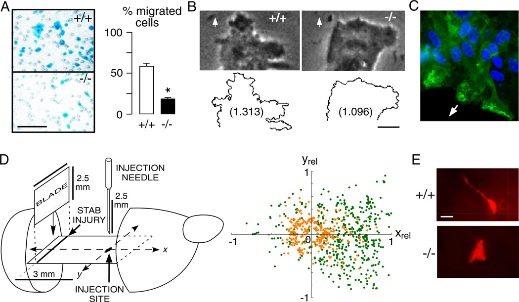
AQP4 facilitates astroglial migration in vitro and in brain. (A) Left. Boyden chamber migration assay showing AQP4+/+ and AQP4−/− astroglia (blue) after scraping off the non-migrated cells. Astroglia were plated on the top chamber of porous transwell filter (2.8 × 10⁴/cm2) and were allowed to migrate for 6 h towards 10% FBS as chemoattractant. Bar, 100 µm. Right. Summary of migration experiments (SEM, * P < 0.001). (B) Phase contrast micrographs (Top) and outline (Bottom) of the leading end of a migrating AQP4+/+ and AQP4−/− astroglial cell in the in vitro wound assay. Arrows show direction of migration. Numbers are fractal dimensions (larger number denotes more irregular cell membrane). Bar 10 µm. (C) AQP4 protein (green) polarization to the front end of migrating astroglia in a wound assay. Arrow shows direction of migration. (D) Left. Stab injury/cell injection model of astroglial migration in mouse brain. Two days before cell injection, a stab was created as shown. Cultured AQP4+/+ and AQP4+/+ astroglia were fluorescently labelled and injected as indicated. Right. Locations of migrating fluorescently stained AQP4+/+ (green) and AQP4−/− (orange) astroglia. The x-axis is relative distance between injection and stab injury sites (xrel). (E) High magnification fluorescence micrographs of migrating AQP4+/+ and AQP4−/− astroglia. Bar 5 µm. Data from Saadoun et al., 2005b and Auguste et al., 2007. (See Color Plate 46.3 in color plate section.)

