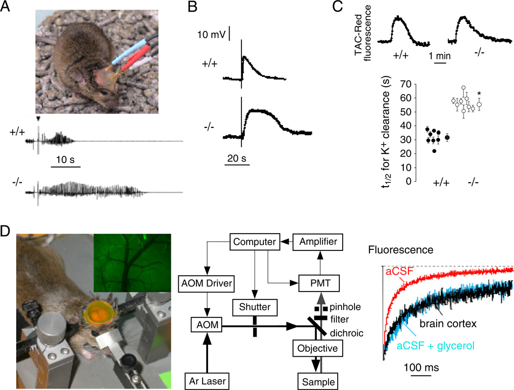
Fig. 4.
AQP4 involvement in brain neuroexcitation. (A) Increased seizure duration in AQP4 null mice. Top. Bipolar electrodes implanted in the right hippocampus were connected to a stimulator and electroencephalograph recording system. Bottom. Representative electroencephalograms in freely moving mice following electrically induced generalized seizures. (B) Delayed K+ clearance in brain following electrically induced seizure-like neuroexcitation. Measurements done using K+-sensitive microelectrodes inserted into brain cortex in living mice. (C) Slowed K+ clearance in brain ECS during cortical spreading depression measured using TAC-Red, a K+-sensitive fluorescent probe. Top. Representative data. Bottom. Half-times (t1/2) for K+ reuptake. (D) Expanded brain ECS in AQP4 null mice measured by cortical surface photobleaching. Left. Mouse brain surface exposed to FITC-dextran with dura intact following craniectomy and fluorescence imaging of cortical surface after loading with FITC-dextran (inset). Middle. Photobleaching apparatus. A laser beam is modulated by an acousto-optic modulator and directed onto the surface of the cortex using a dichroic mirror and objective lens. Right. In vivo fluorescence recovery in cortex of wildtype mouse shown in comparison to aCSF and 30% glycerol in aCSF. Taken from Binder et al., 2006, Binder et al., 2004b and Padmawar et al., 2005. (See Color Plate 46.4 in color plate section.)

