Figure 5.
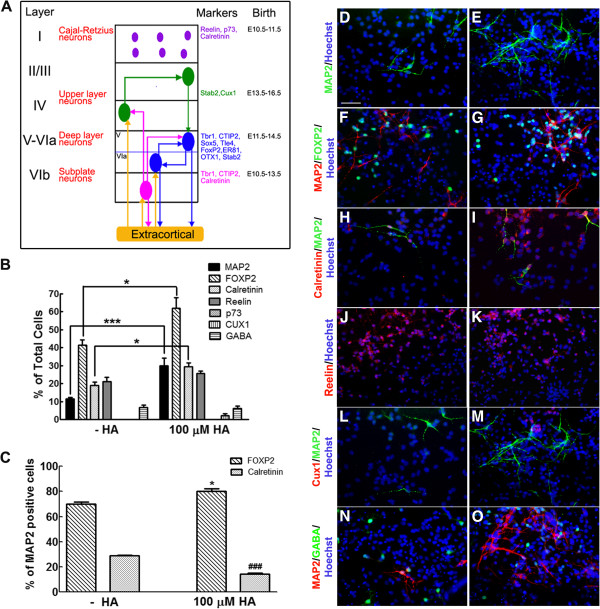
HA induces differentiation of NPC to FOXP2 neurons. (A) Modified scheme illustrating the expression of layer-specific markers in cortical neurons, their timing of generation in vivo and regions of projection [8,21,22]. (B) Quantification of the total number of mature neurons (MAP2+) and detection of specific cortical laminar neuronal phenotypes. *P <0.05 and ***P <0.001. (C) Percentage of MAP2-positive neurons that express FOXP2 or calretinin. Note that the proportion of FOXP2+ neurons increased whereas calretinin+ neurons decreased after HA treatment. *P <0.05 versus FOXP2 without HA; ###P <0.001 versus calretinin without HA. (D-O) Representative micrographs of immunofluorescence detection of the indicated markers in control (D, F, H, J, L and N) and HA-treated (E, G, I, K, M and O) cells. Nuclei were labeled with Hoechst and are shown in blue. Scale bar = 100 μm. HA, histamine; MAP2, microtubule associated protein 2; NPC, neural precursor cells.

