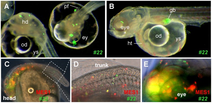
Figure 4. Pluripotency in vivo.
Embryos at the midblastula stage were transplanted with ES cells and photographed at day 3 (A) and 7 post fertilization (B–E). (A and B) Merged micrographs of chimeras, showing GFP-labeled ES cells of p53-targeted clone #22 in many compartments and organ systems including the eye (ey), head (hd), heart (ht), gall bladder (gb) and pectoral fin (pf). od, oil droplet; ys, yolk sac. (C–E) Co-distribution of parental MES1 and p53-targeted clone #22 in the developing chimera at day 7 post fertilization. Following genetic labeling by plasmid transfection, parental MES1 (red) and clone #22 (green) were mixed at 1∶1 ratio and transplanted at approximately 200 cells per blastula. (D and E) Larger magnification of the posterior trunk framed in (C) and anterior end. The embryo is 1 mm in diameter.

