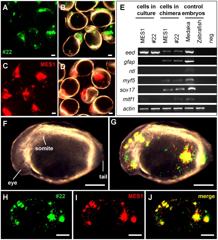
Figure 5. Chimeric assay of pluripotency in vivo.
Zebrafish blastulae were used as the host for transplantation of MES1 (red), #22 (green) or both and analyzed at 1 dpf by microscopy and RT-PCR. (A and B) Chimeras by transplantation of #22 cells on fluorescent micrograph (A) and merge between fluorescent and brightfield optics (B). (C and D) Chimeras by transplantation of MES1 cells on fluorescent micrograph (C) and merge between fluorescent and brightfield optics (D). (E) RT-PCR analysis of gene expression. MES1, #22, 1-day-old embryos of medaka and zebrafish were used for comparison. Primers used were specific to medaka cDNAs, except for β-actin primers that amplify the β-actin cDNA of both medaka and zebrafish. (F–J) Chimera by cotransplantation of MES1 and #22 cells on brightfield (F), brightfield-fluorescent merge (G), GFP fluorescence (H), RFP fluorescence (I) and merge between GFP and RFP optics (J). Scale bars, 200 µm.

